

")


Volume 3, Nº 3 (2022)
- Ano: 2022
- ##issue.datePublished##: 17.10.2022
- Artigos: 7
- URL: https://jdigitaldiagnostics.com/DD/issue/view/3845
- DOI: https://doi.org/10.17816/DD.33

Technical Reports
Optimized biparametric magnetic resonance imaging protocol for prostate cancer detection
Resumo
BACKGROUND: Prostate cancer is one of the most commonly diagnosed cancers in men worldwide. PI-RADS v2.1 contains the requirements for the magnetic resonance imaging protocol, which cannot be fully implemented on a significant component of functioning scanners. Consequently, magnetic resonance imaging approaches vary in different medical organizations and often do not allow for a qualitative interpretation of images and diagnosis of the target pathology.
AIM: To develop a biparametric magnetic resonance imaging protocol optimized for the existing magnetic resonance imaging scanners for the diagnosis of prostate cancer and to allow the screening and detection of neoplasms as early as possible. Simultaneously, the protocol should fulfill the current PI-RADS v2.1 recommendations to the maximum possible extent and meet the requirements of effective workflow in the radiology department.
MATERIALS AND METHODS: Preliminary analysis of prostate magnetic resonance imaging scanning in medical organizations of the Moscow Health Care Department showed the absence of a unified approach. Using the iterative adjustment of scanning parameters, we adjusted the protocol to ensure acceptable quality with maximum available compliance with PI-RADS v2.1.
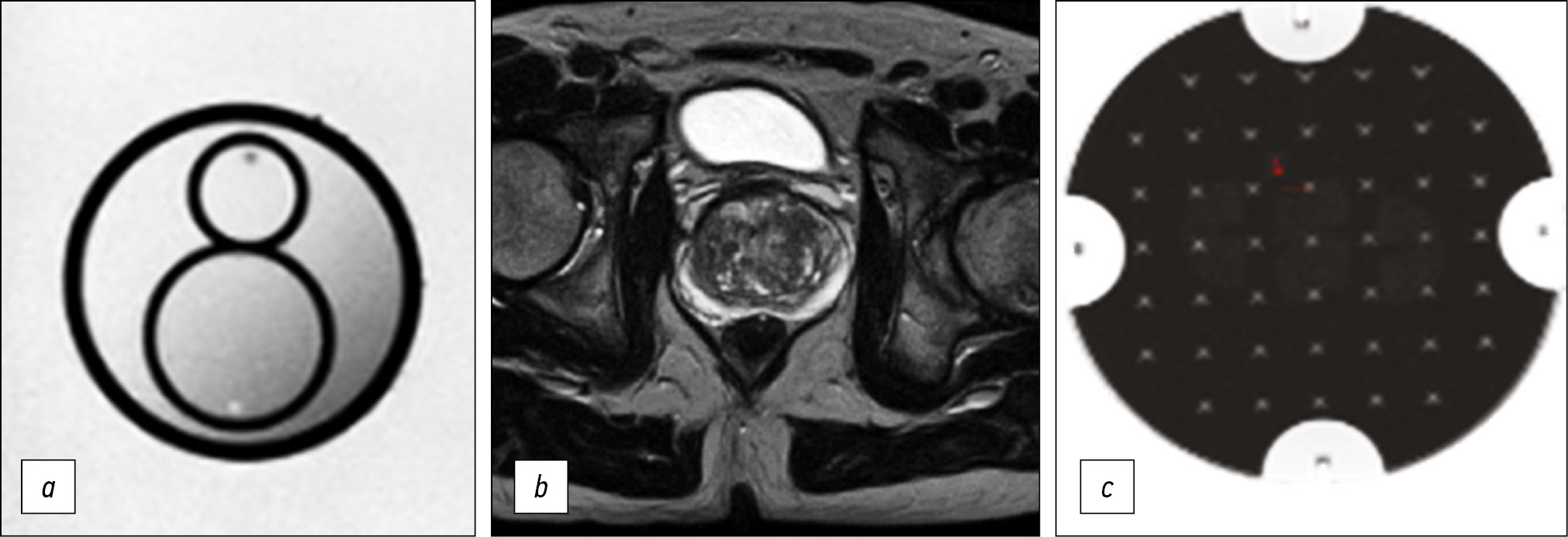
To quantify the quality of the images, we used the magnetic resonance imaging phantom recommended by the American College of Radiology.
RESULTS: The biparametric protocol was developed for Excelart Vantage 1.5 T, including T2-weighted images in three planes and diffusion-weighted images, which took less than 11 min. Moreover, the image quality parameters (intensity inhomogeneity, nonlinearity, resolution, and slice thickness) were within the acceptable ranges recommended by the magnetic resonance imaging manufacturer.
CONCLUSION: The prostate may be effectively evaluated using the proposed magnetic resonance imaging protocol. Introducing it into practice could have a significant impact on the detection of prostate cancer in men. The entire duration of the protocol provides a possibility to supplement it with any sequences, depending on the final purpose of investigation.
 166-177


Reviews
Evolution of research and development in the field of artificial intelligence technologies for healthcare in the Russian Federation: results of 2021
Resumo
The use of artificial intelligence technologies in Russian healthcare is a priority area for implementing a national strategy for the development of artificial intelligence in the country. The introduction of digital solutions based on artificial intelligence in healthcare facilities should improve the standard of living of the population and the quality of medical care, including areas of preventive examinations, diagnostics based on image analysis, prediction of disease development, selection of optimal drug dosages, reducing the threat of pandemics, and automating and increasing the accuracy of surgical interventions.
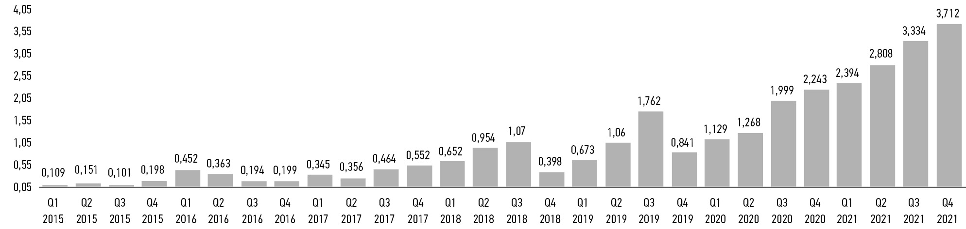
Policy management and technical regulation are under development in the field of artificial intelligence in healthcare. The domestic market for relevant solutions has been created, and some products have been certified as medical devices from Roszdravnadzor (Federal Service for Surveillance in Healthcare). Various teams of scientists are conducting research. However, Russia is still behind the leading countries in the field of artificial intelligence, such as the United States and China. Investments in healthcare products based on artificial intelligence decreased significantly in 2021. The major reasons for the lag, at least in terms of market indicators, are low demand and the inability of state medical organizations to fund artificial intelligence projects. There are also other issues related to trust in the safety and effectiveness of such solutions.
178-194
Sarcopenia: modern approaches to solving diagnosis problems

Resumo
Although sarcopenia is a relatively new diagnosis for medical statistics and the healthcare system, it represents a social and economic burden on the healthcare due to the large number of possible adverse outcomes such as increased risk of falls, physical disability, longer hospital stays, and increased mortality. No specialized medical treatment is available for sarcopenia; however, prevention and timely nonpharmacological treatment can reduce the risk of potential adverse effects. To establish the diagnosis of sarcopenia, it is necessary to confirm the decrease in not only muscle strength but also muscle mass. Instrumental diagnostics includes methods such as dual-energy X-ray absorptiometry and bioimpedance analysis. These methods can be supplemented by artificial intelligence algorithms for the automatic segmentation of muscle and fat tissue on computed tomography and magnetic resonance images, followed by calculation of the skeletal muscle index at the level of the L3 vertebra (L3SMI). Such software, when used in systems such as the Unified Radiological Information Service of the Unified Medical Information and Analytical System of Moscow, opens up opportunities for opportunistic screening. However, despite the recognition of CT and MRI as the “gold standard” by the European Working Group on Sarcopenia in Older People, there are no generally accepted L3SMI cut-off values for CT and MR diagnostics of sarcopenia. Furthermore, there is the problem of unifying the term “skeletal muscle index.” If these problems could be solved through further population studies, it will be possible to obtain a new method for the instrumental diagnosis of sarcopenia with its subsequent use for opportunistic screening.
196-211
Patient dose monitoring software in radiology
Resumo
An increase in the number of diagnostic procedures using ionizing radiation (computed tomography, interventional procedures, and the use of nuclear medicine) results in an increase in radiation exposure and, consequently, an increase in collective and individual doses of radiation to patients.
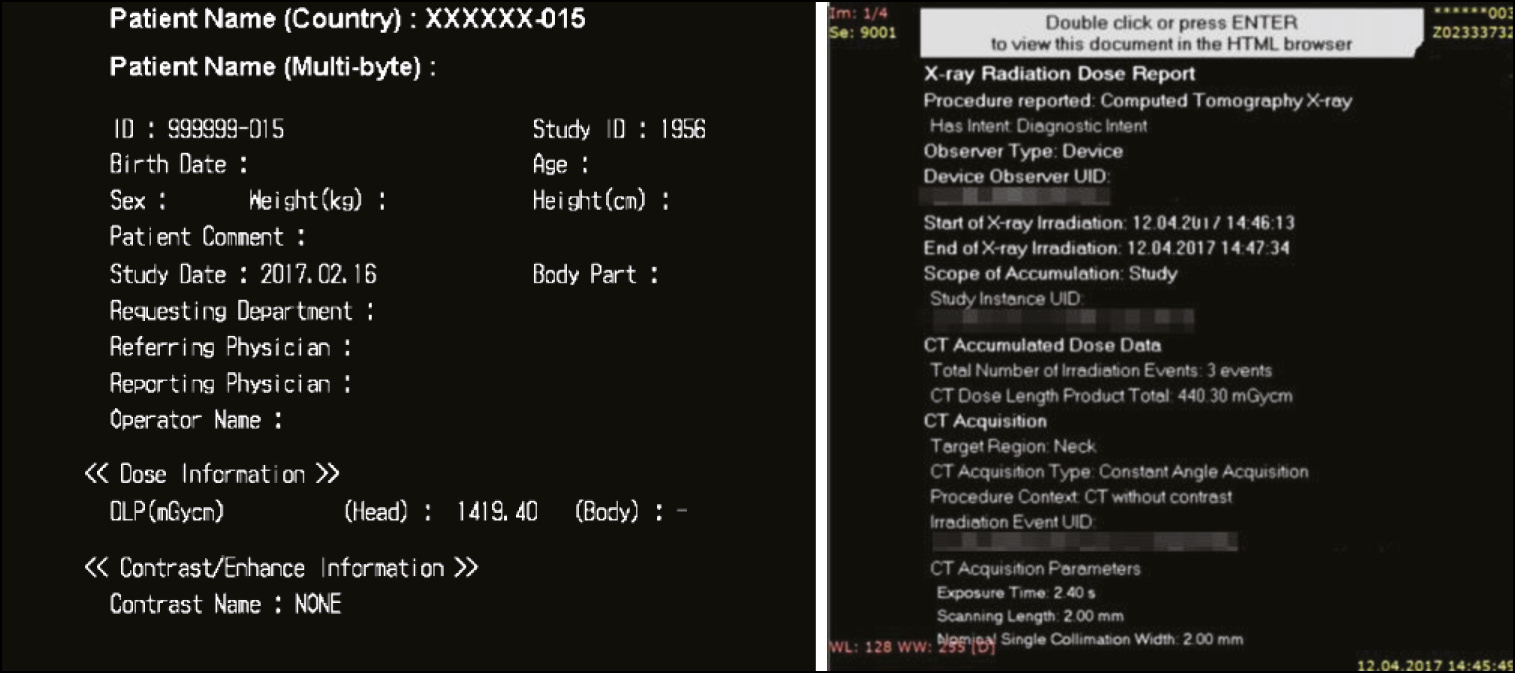
Diagnostic studies from the international professional community are extensively focusing on issues such as management and dose optimization. Worldwide practice can resolve these issues using software for monitoring patient doses to automatically collect, analyze, and account for patient doses in various types of diagnostic studies. The software allows to obtain data on the doses of patients from X-ray procedures and detailed information about studies, track the total accumulated dose of the patient, and maintain statistics on the device, X-ray laboratory, and the medical organization. It also helps analyze the collected dosimetric data, deduce the causal relationship between dose indications and diagnostic procedure conditions, and monitor the effectiveness of the equipment.
The basic capabilities of patient dose monitoring software (DMS) available on the global market were investigated. The major technical requirements for the software functional needed in practical work were defined.
Modern DMS have a wide range of possibilities for automated collection, storage, and management of patient radiation exposure data in radiology departments. DMS increase the quality of healthcare services, provide patient safety, and optimize the workflow of medical organizations.
212-230
Transparent Reporting of a multivariable prediction model for Individual Prognosis Or Diagnosis (TRIPOD): Explanation and Elaboration. Translation in to Russian
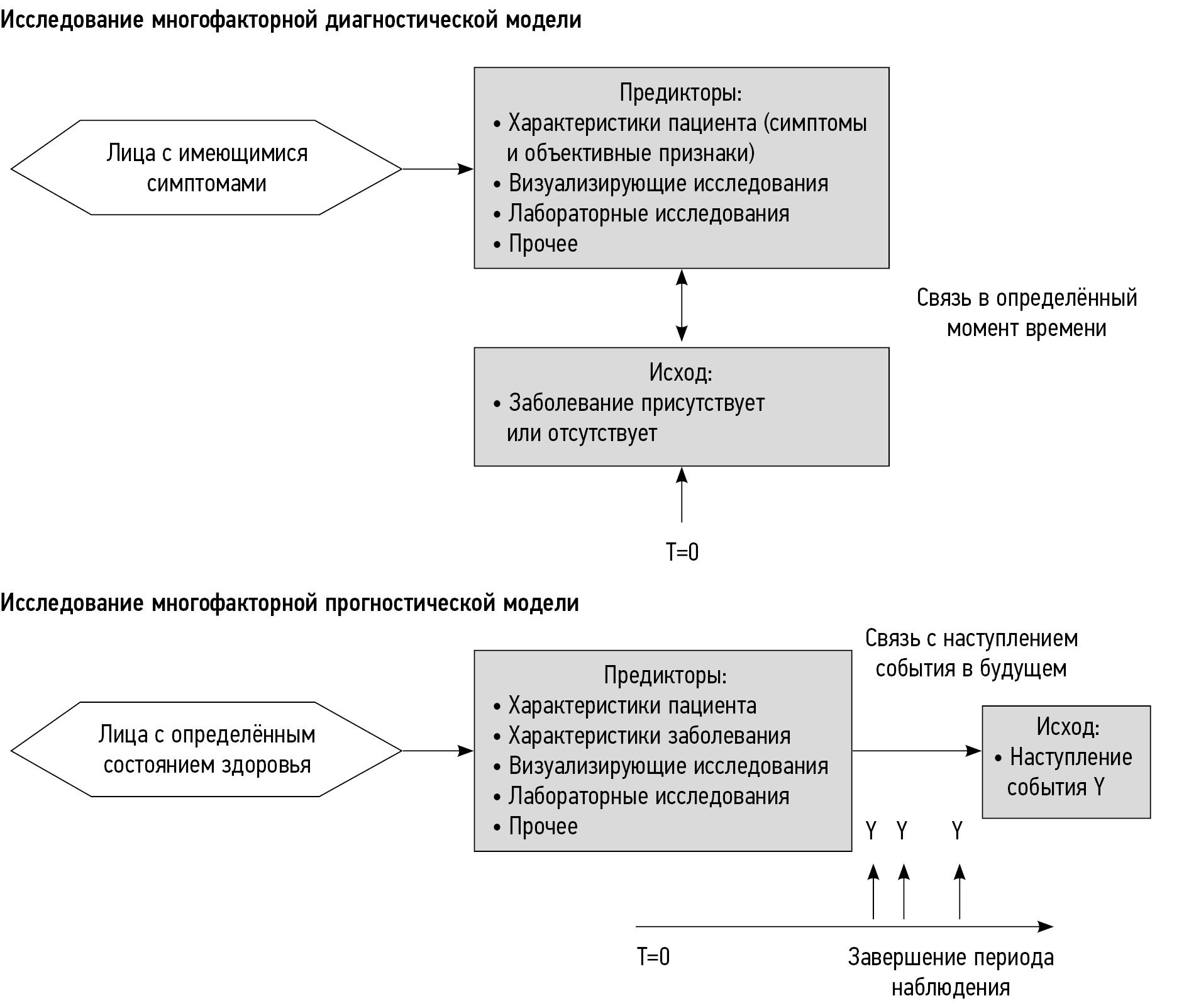
Resumo
The TRIPOD (Transparent Reporting of a multivariable prediction model for Individual Prognosis Or Diagnosis) Statement includes a 22-item checklist, which aims to improve the reporting of studies developing, validating, or updating a prediction model, whether for diagnostic or prognostic purposes. The TRIPOD Statement aims to improve the transparency of the reporting of a prediction model study regardless of the study methods used. This explanation and elaboration document describes the rationale; clarifies the meaning of each item; and discusses why transparent reporting is important, with a view to assessing risk of bias and clinical usefulness of the prediction model. Each checklist item of the TRIPOD Statement is explained in detail and accompanied by published examples of good reporting. The document also provides a valuable reference of issues to consider when designing, conducting, and analyzing prediction model studies. To aid the editorial process and help peer reviewers and, ultimately, readers and systematic reviewers of prediction model studies, it is recommended that authors include a completed checklist in their submission. The TRIPOD checklist can also be downloaded from www.tripod-statement.org.
For members of the TRIPOD Group, see the Appendix.
This article is the translation in to Russian by Dr. Ruslan Saygitov (ORCID: 0000-0002-8915-6153) from the original published in [Ann Intern Med. 2015; 162:W1-W73. doi: 10.7326/M14-0698 ].
232-322
Correspondence
Coexistence of machine intelligence, cyber art, and diagnostics: is it possible?
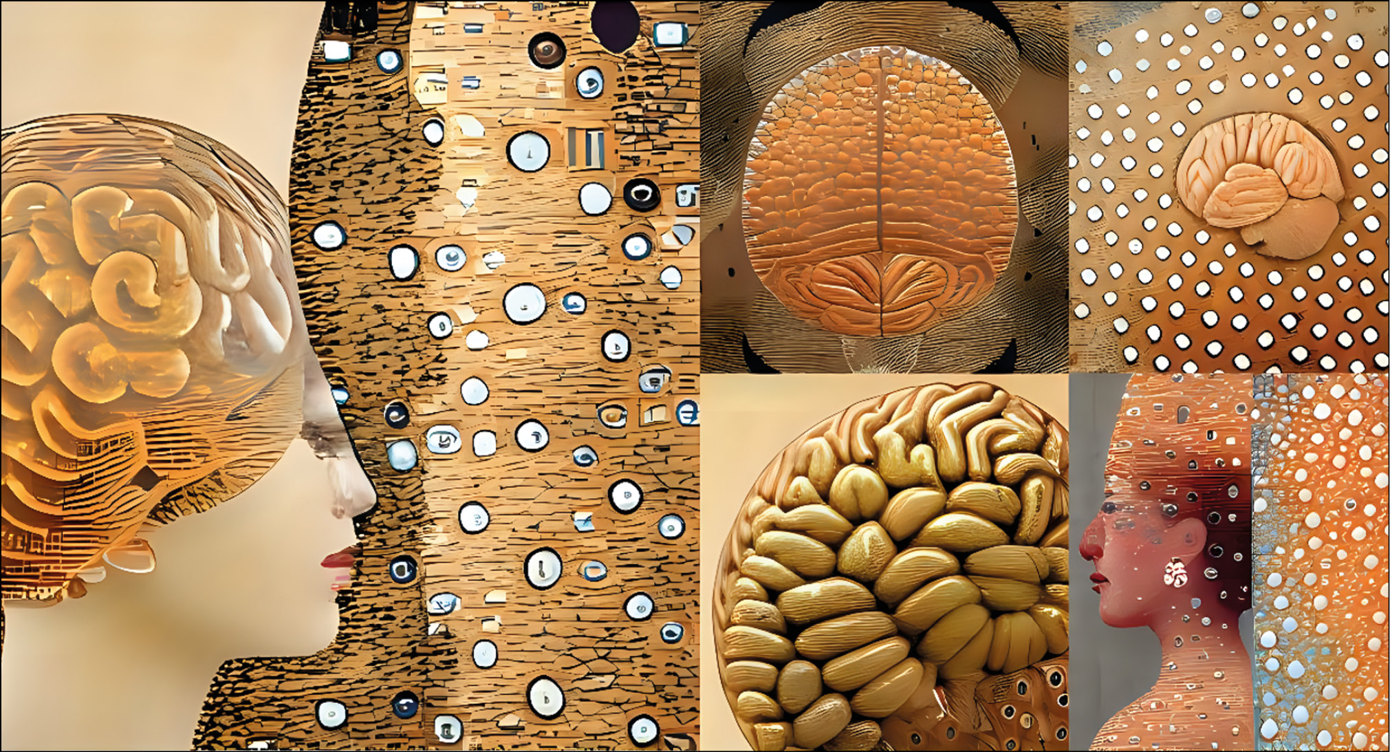
Resumo
The development of machine intelligence and the application of generative images created using it is a promising area of communication design and human–machine interaction. This letter to the editor represents the author’s vision of the use of generative images for diagnosing human conditions.
The use of machine intelligence as an interactive and intelligent diagnostic tool will allow a psychologist and a physician to effectively complement the therapeutic processes of controlled interactions of their users.
Libraries of models and sets of applications with text-to-image algorithms are already available that can be used by engineers and designers in the process of creating objects of modern digital art. They can also be applied in the investigation of new paradigms using visual communications and their application in experimental diagnostics.
324-330
Case reports
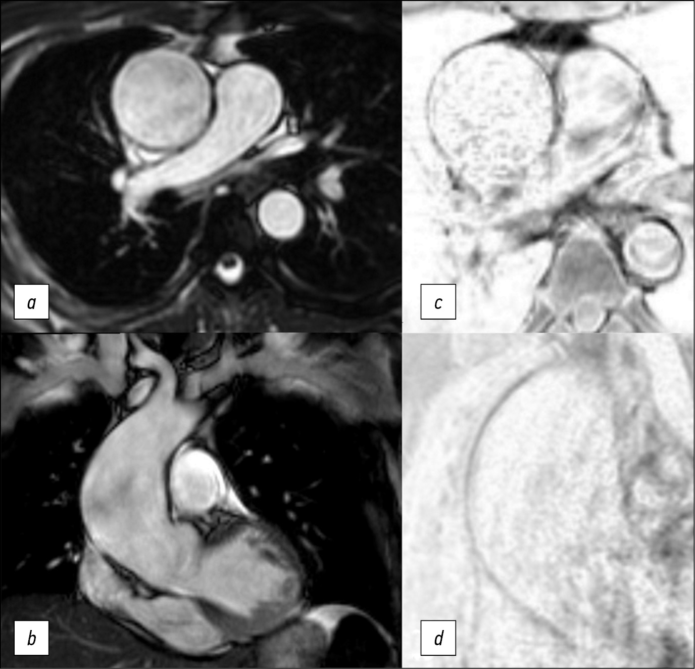
Diagnostic challenge: innovative approach in use of magnetic resonance imaging in aortic aneurysm
Resumo
Here we report a case of technological innovation: the use of magnetic resonance imaging to determine surgical strategy.
Here is a 47-year-old man who underwent an magnetic resonance imaging and subsequent surgical treatment of the aortic aneurysm. Unlike echocardiography, magnetic resonance imaging enabled us to view the entire thoracic aorta. Unlike computer tomography, magnetic resonance imaging enabled us to detect changes in the aortic wall accurately. Thus, in this case, the use of magnetic resonance imaging allowed us to determine the distal resection edge. The patient`s postoperative course was unremarkable. Use of electrocardiogram-synchronized magnetic resonance imaging of thoracic aorta allows detecting structural changes of the aortic wall and its mechanical properties. It is significant that magnetic resonance imaging results of the aortic wall correlate with histologic examination.
The extent of changes in the aortic wall must be determined to accurately plan surgical treatment of patients with aortic aneurism.
Magnetic resonance imaging of the aortic wall is promising for further study in multicenter research.
332-339
Регистрационный номер и дата принятия решения о регистрации СМИ: серия ПИ № ФС 77 - 79539 от 09 ноября 2020 г.