

")


Volume 2, Nº 2 (2021)
- Ano: 2021
- ##issue.datePublished##: 10.08.2021
- Artigos: 8
- URL: https://jdigitaldiagnostics.com/DD/issue/view/3395
- DOI: https://doi.org/10.17816/DD.22

Original Study Articles
Opportunities to reduce the radiation exposure during computed tomography to assess the changes in the lungs in patients with COVID-19: use of adaptive statistical iterative reconstruction
Resumo
BACKGROUND: Several COVID-19 patients are subjected to multiple imaging examinations during hospitalization, the cumulative effect of which can significantly increase the total dose of radiation received. The effective radiation dose can be reduced by lowering the current and voltage of the X-ray tube, but this reduces image quality. One possible solution is to use adaptive statistical iterative reconstruction technology on the «raw» CT data. Recently, data on the efficacy of low-dose CT (LDCT) in the diagnosis of COVID-19 have appeared in the literature.
AIM: To analyze the quality and diagnostic value of LDCT images of the lungs after applying an iterative processing algorithm and to assess the possibility of reducing the radiation load on the patient when diagnosing COVID-19.
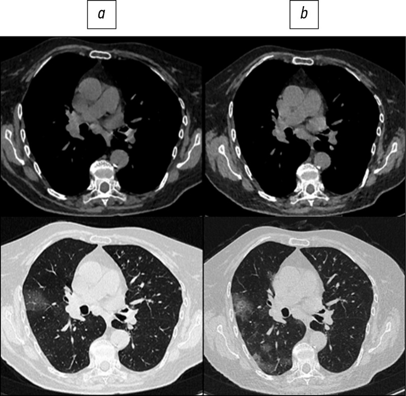
MATERIALS AND METHODS: Patients from the Infectious Diseases Department of the Moscow State University Hospital participated in the prospective study. CT examinations were performed at the time of patient admission and discharge and were repeated as needed during hospitalization. In the first study, a standard CT protocol with a tube voltage of 120 kV and automatic current modulation in the range of 200–400 mA was used; in repeated CT scans, the LDCT protocol was used with reduced tube voltage parameters (100 or 110 kV) and automatic current modulation in the range of 40–120 mA. To assess the diagnostic value of LDCT in comparison with standard CT, a survey was conducted among doctors from the Department of Radiation Diagnostics at Moscow State University Hospital. The questionnaire included a comparison of the two methods for identifying the following pathological processes: «ground-glass» opacities, compaction of the lung tissue with reticular changes, areas of lung tissue consolidation, and lymphadenopathy.
RESULTS: The study included 151 patients. The average age was 58±14.2 years, with men accounting for 53.6% of the population. During LDCT the radiation load was reduced by 2.96 times on average, CTDI by 2.6 times, DLP by 3.1 times, the current on the tube by 1.83 times, and the voltage on the tube by 1.2 times. The results indicate that the effectiveness of detecting the main signs of viral pneumonia and assessing the dynamics of the patient’s condition does not differ significantly from CT performed according to the standard protocol.
CONCLUSIONS: The results of a comparison of standard and low-dose CT show that there is no significant loss of diagnostic information and image quality as the radiation load is reduced. Thus, chest LDCT can be used to successfully diagnose COVID-19 in routine practice.
 94-104


Inter-observer variability between readers of CT images: all for one and one for all
Resumo
BACKGROUND: The markup of medical image datasets is based on the subjective interpretation of the observed entities by radiologists. There is currently no widely accepted protocol for determining ground truth based on radiologists’ reports.
AIM: To assess the accuracy of radiologist interpretations and their agreement for the publicly available dataset “CTLungCa-500”, as well as the relationship between these parameters and the number of independent readers of CT scans.
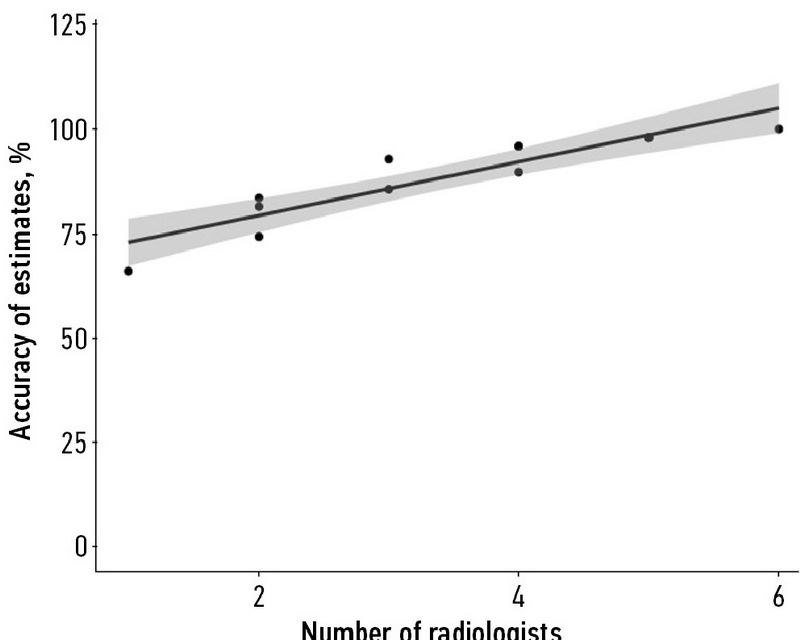
MATERIALS AND METHODS: Thirty-four radiologists took part in the dataset markup. The dataset included 536 patients who were at high risk of developing lung cancer. For each scan, six radiologists worked independently to create a report. After that, an arbitrator reviewed the lesions discovered by them. The number of true-positive, false-positive, true-negative, and false-negative findings was calculated for each reader to assess diagnostic accuracy. Further, the inter-observer variability was analyzed using the percentage agreement metric.
RESULTS: An increase in the number of independent readers providing CT scan interpretations leads to accuracy increase associated with a decrease in agreement. The majority of disagreements were associated with the presence of a lung nodule in a specific site of the CT scan.
CONCLUSION: If arbitration is provided, an increase in the number of independent initial readers can improve their combined accuracy. The experience and diagnostic accuracy of individual readers have no bearing on the quality of a crowd-tagging annotation. At four independent readings per CT scan, the optimal balance of markup accuracy and cost was achieved.
105-118
Reviews
Strengthening the Reporting of Observational Studies in Epidemiology (STROBE): Explanation and Elaboration. Translation to Russian
Resumo
Much medical research is observational. The reporting of observational studies is often of insufficient quality. Poor reporting hampers the assessment of the strengths and weaknesses of a study and the generalisability of its results. Taking into account empirical evidence and theoretical considerations, a group of methodologists, researchers, and editors developed the Strengthening the Reporting of Observational Studies in Epidemiology (STROBE) recommendations to improve the quality of reporting of observational studies. The STROBE Statement consists of a checklist of 22 items, which relate to the title, abstract, introduction, methods, results and discussion sections of articles. Eighteen items are common to cohort studies, case-control studies and cross-sectional studies and four are specific to each of the three study designs. The STROBE Statement provides guidance to authors about how to improve the reporting of observational studies and facilitates critical appraisal and interpretation of studies by reviewers, journal editors and readers. This explanatory and elaboration document is intended to enhance the use, understanding, and dissemination of the STROBE Statement. The meaning and rationale for each checklist item are presented. For each item, one or several published examples and, where possible, references to relevant empirical studies and methodological literature are provided. Examples of useful flow diagrams are also included. The STROBE Statement, this document, and the associated Web site (http://www. strobe-statement.org/) should be helpful resources to improve reporting of observational research.
This article is the reprint with Russian translation of the original that can be observed here: Vandenbroucke JP, von Elm E, Altman DG, Gotzsche PC, Mulrow CD, et al. Strengthening the Reporting of Observational Studies in Epidemiology (STROBE): Explanation and Elaboration. PLoS Med. 2007;4(10):e297. doi: 10.1371/journal.pmed.0040297.
119-169
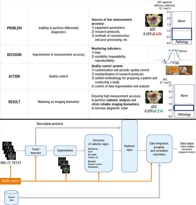
The role of the quality control system for diagnostics of oncological diseases in radiomics
Resumo
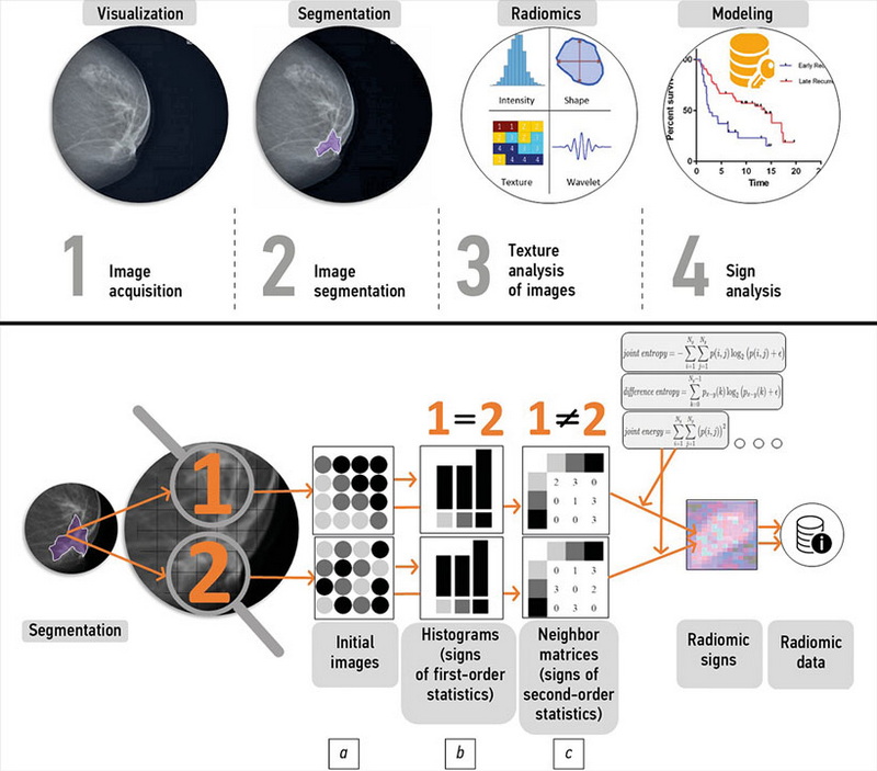
Modern medical imaging methods allow for both qualitative and quantitative evaluations of tumors and issues surrounding them. Advances in computer science and big data processing are transforming any radiological study into analytic datasets, especially with the use of machine learning in medical image analysis. Among these datasets, statistically significant correlations with clinical events can then be searched for to subsequently assess their predictive value and ability to predict a particular clinical outcome. This concept, known as “radiomics,” was first described in 2012. It is particularly important in oncology because each type of tumor can be subdivided into many different molecular genetic subtypes, and simple visual characteristics are no longer sufficient. Moreover, as an absolutely noninvasive method, radiomics can provide a radiologist with additional information that would otherwise be unavailable without a histological examination of biopsy material. However, as with any methodology based on the use of big data, the question of the quality of the initial data becomes critical, because this can directly affect the outcome of the analysis and provide incorrect diagnostic information.
In this literature review, we examine potential approaches to ensuring the quality of research at all stages, from technical control of the state of diagnostic equipment to the extraction of imaging markers in oncology and the calculation of their correlation with clinical data.
170-184
The role of mammography in breast cancer radiomics
Resumo
Mammography is still the only screening method for breast cancer. Although digital mammography is the most common and widely used method for detecting breast cancer, it is ineffective at detecting and assessing intratumoral heterogeneity. Due to the small size of the tissue sample or tumor, biopsies often fail to represent the entire tumor. For this reason, selecting a treatment and determining a patient’s prognosis becomes difficult. In this case, medical imaging is a noninvasive approach that can provide a more comprehensive view of the entire tumor, act as a “virtual biopsy,” and be useful for monitoring disease progression and response to therapy.
Radiomics with texture analysis allows you to look at an image as a group of numerical data, moving beyond the usual visual perception and into a deeper analysis of digital, pixel data to improve the accuracy of differential diagnosis. Radiogenomics is a natural extension of radiomics that focuses on determining gene expression based on radiologic tumor phenotype. The purpose of this review is to evaluate the role of mammography in breast cancer radiomics and radiogenomics.
The article presents a literature review of relevant Russian scientific articles found in databases such as PubMed, Medline, Springer, eLibrary, and Google Scholar. The information obtained was then pooled, structured, and analyzed to examine the role of mammography in breast cancer screening radiomics.
185-199
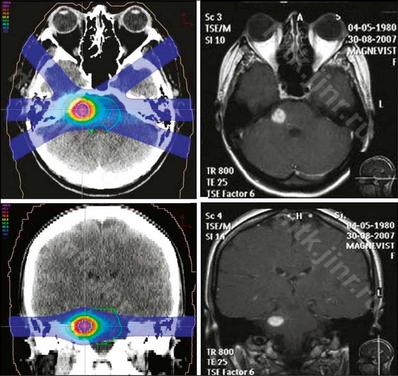
Cavernous malformations of the brain and modern views on their treatment
Resumo
Cavernous malformations of the brain have become an increasingly common pathology in recent years, thanks to the advancement of modern methods of neuroimaging. Despite the benign nature of the course in most cases, these formations can cause convulsions and serious neurological disorders. Typically, clinical manifestations are caused by hemorrhages in the structure of the cavernous and surrounding parenchyma of the brain. The management strategy chosen for patients with cerebral cavernous malformations is determined by the type of malformation, its size, localization, the presence of repeated hemorrhages, and the clinical picture.
This literature review focuses on modern methods of treating cerebral cavernous malformations. The main methods of treatment for cavernous malformations of the brain, particularly surgical treatment, have been analyzed. If surgical intervention is not possible, alternative methods of treatment include radiation therapy, such as stereotaxic radiosurgery, and proton therapy, in cases of deep location of foci in functionally significant areas of the brain, which are characterized by the highest risk of complications. Thе possibilities, efficacy, and safety of stereotactic radiosurgical treatment are discussed, as well as the use of proton therapy in the treatment of cavernous malformations. Furthermore, radiation therapy has been shown to be beneficial for cavernous malformations.
200-210
Fatigue in radiology: a fertile area for future research

Resumo
Fatigue in radiologists may be responsible for a large number of medical errors. This review describes the latest research on fatigue in radiology. This includes measurement methods, and recent evidence on how fatigue affects accuracy in laboratory test conditions and in clinical practice. The extensive opportunities for future research in the area are explored, including testing interventions to reduce fatigue-related error, and further understanding of which fatigue measures correlate with errors. Finally we explore the possibility of answering these questions using large population-based observational studies and pragmatic integrated randomised controlled trials.
This publication is the reprint with Russian translation from original: Taylor-Phillips S, Stinton C. Fatigue in radiology: a fertile area for future research. Br J Radiol. 2019;92:20190043. doi: 10.1259/bjr.20190043.
211-222
Case reports
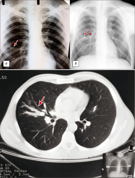
Long-term broncocele anamnesis, triggered by typical carcinoid
Resumo
The paper presents a case of a single bronchocele (bronchogenic retention cyst) caused by a typical carcinoid that was observed for a long time. During the initial complex examination, including computed tomography with intravenous contrast, fibrobronchoscopy, and immunological and bacteriological examinations of tuberculosis, there were no changes for the oncological and infectious nature. The changes were interpreted as the result of a postponed nonspecific inflammatory process. Most of them were monitored using chest X-ray and the changes were stable. After 15 years, a control chest X-ray revealed an increase in the size of the compaction in the lung and the appearance of a mass with calcification in the medial sections of the compaction zone. Additional examination, including computed tomography with biopsy, determined that the obstruction of the bronchus was caused by a neoplasm [according to histological examination (typical carcinoid)].
It should be noted that the initial detection of negative study results requires oncological alertness and periodic examinations in dynamics.
223-230
Регистрационный номер и дата принятия решения о регистрации СМИ: серия ПИ № ФС 77 - 79539 от 09 ноября 2020 г.