

")


Automated radiology workstation: comparing two approaches for selecting radiodiagnostic solutions (technical report)
- Authors: Vasilev Y.A.1,2, Slavushcheva E.A.1, Zelenova M.A.1, Shulkin I.M.1, Arzamasov K.M.1,3
-
Affiliations:
- Research and Practical Clinical Center for Diagnostics and Telemedicine Technologies
- National Medical and Surgical Center named after N.I. Pirogov
- MIREA — Russian Technological University
- Issue: Vol 6, No 3 (2025)
- Pages: 477-486
- Section: Technical Reports
- Submitted: 02.05.2024
- Accepted: 20.03.2025
- Published: 14.10.2025
- URL: https://jdigitaldiagnostics.com/DD/article/view/631519
- DOI: https://doi.org/10.17816/DD631519
- EDN: https://elibrary.ru/FDMNZG
- ID: 631519

Cite item
Full Text

Abstract
BACKGROUND: Patient examinations generate large amounts of data on various diseases, which are virtually impossible to process manually. Currently, automated radiology workstations incorporate clinical decision–support systems that facilitate image analysis. At present, two options are used: built-in vendor-dependent and vendor-independent software solutions. Both have broad functionality, but they also have their advantages and drawbacks. Therefore, the choice of the best clinical decision–support system depends on the healthcare organization.
AIM: This study aimed to compare two approaches for selecting radiodiagnostic solutions for automated radiology workstations in Moscow.
METHODS: The study conducted a two-stage, cross-sectional survey between September 2023 and March 2024. Data on vendor-independent software were obtained from participants of the Experiment on the use of innovative computer vision technologies for analysis of medical images in the Moscow healthcare system. Conversely, data on vendor-dependent solutions was collected from the manufacturers’ websites. Furthermore, a survey of 40 radiologists was performed to evaluate the relevance of software functions.
RESULTS: At the time of the survey, the vendor-independent software demonstrated slightly more functions than the vendor-dependent solutions. The greatest differences were observed in the functionality of computed tomography and magnetic resonance imaging modalities, whereas the functionality for X-ray imaging and mammography was nearly identical. The survey of radiologists showed that 6 of 17 functions were unique to built-in vendor-dependent software. However, <40% of the radiologists actually needed these functions, whereas the shared functions were relevant for >50% of the respondents.
CONCLUSION: Vendor-independent and built-in vendor-dependent software solutions share only half of their functions. Thus, in choosing between these two options, healthcare facilities should consider their specialization and their radiologists’ requests. Moreover, approximately two-thirds of the built-in vendor-dependent software functions used by radiologists in Moscow can be implemented using vendor-independent solutions. Therefore, the choice of software should be based on the facility’s technical capacity and economic feasibility.
Full Text
ОБОСНОВАНИЕ
Современную медицинскую практику невозможно представить без информационных технологий. Рост числа пациентов обусловливает высокую нагрузку как на систему здравоохранения в целом, так и на отдельные учреждения. При этом накопление больших массивов данных о различных заболеваниях делает их ручную обработку невозможной [1, 2]. В связи с этим на автоматизированных рабочих местах (АРМ) врачей-рентгенологов всё активнее применяют системы поддержки принятия врачебных решений, обеспечивающие анализ медицинских изображений [3–5].
Наиболее распространёнными являются следующие два подхода к организации АРМ врача-рентгенолога:
- сиспользованием готового решения отпроизводителя рентгеновского оборудования — встроенное вендор-зависимое программное обеспечение (ПО);
- ссамостоятельным подбором компонентов АРМ — вендор-независимое ПО1.
Оба подхода возможно применять для:
- получения независимого мнения — когда врач видит результаты работы ПО только после того, как закончил работать над описанием исследования;
- параллельного прочтения — врач видит результаты работы ПО одновременно с изображением, полученным с диагностического оборудования;
- приоритизации исследований — исследования, оценённые ПО как патологические или срочные, поступают врачу на интерпретацию первыми;
- их исключения — исследования, оценённые ПО как непатологические, поступают на оценку врачу в порядке общей очереди.
Вендор-зависимые и вендор-независимые решения обладают широким функционалом и продолжают совершенствоваться за счёт применения технологий искусственного интеллекта [6]. В связи с этим перед руководителями в сфере организации здравоохранения (министрами, главными врачами, их заместителями и др.) закономерно возникает вопрос: какое ПО предпочтительнее для использования в конкретной медицинской организации — вендор-зависимое или вендор-независимое?
ЦЕЛЬ
Сравнить два подхода к применению ПО для анализа лучевых исследований в условиях АРМ врача-рентгенолога на примере Москвы.
МЕТОДЫ
Дизайн работы: двухэтапное одномоментное опросное исследование.
На первом этапе выполнен анализ функций вендор-зависимых и вендор-независимых решений, доступных для использования в Москве. Информация о вендор-независимых решениях получена из каталога сервисов на основе искусственного интеллекта, применяемых в Московском эксперименте по использованию инновационных технологий в области компьютерного зрения для анализа медицинских изображений и дальнейшего применения этих технологий в системе здравоохранения2. Информация о вендор-зависимых решениях агрегирована с сайтов компаний-производителей34567891011121314151617.
На втором этапе исследования проведён социологический опрос на базе московского референсцентра, в котором приняли участие 40 врачей. Для оценки практической значимости основных функций вендор-зависимого ПО мы разработали опросник «Практическая значимость основных функций вендор-зависимого программного обеспечения» (Приложение 1). Опросник мы составили самостоятельно для целей данного исследования, и он не проходил формальную процедуру валидации. Инструмент содержал 17 закрытых вопросов, каждый из которых посвящён оценке одной конкретной функции на основе предыдущего практического опыта респондентов. Значимость функций оценивали по 3-балльной номинальной шкале: «Высокая», «Средняя», «Низкая». Для исключения смещающих факторов по каждому вопросу предусмотрен вариант ответа «Не использовал(-а)/не знаю».
Работа проведена в период с сентября 2023 г. по март 2024 г.
Этическая экспертиза
Данное исследование основано на результатах Эксперимента по использованию инновационных технологий в области компьютерного зрения для анализа медицинских изображений и дальнейшего применения в системе здравоохранения города Москвы, утверждённого этическим комитетом (выписка из протокола № 2 НЭК МРО РОРР от 20.02.2020), также зарегистрированного на ClinicalTrials (NCT04489992).
РЕЗУЛЬТАТЫ
Сравнение функциональности вендор-зависимых и вендор-независимых решений
Число функций, доступных на момент выполнения работы, у вендор-независимого ПО оказалось несколько больше, чем у вендор-зависимого (Приложение 2).
Примечательно, что количество функций вендор-зависимых и вендор-независимых решений для модальности «Рентгенография» оказалось почти одинаковым. Аналогичную картину наблюдали и для модальности «Маммография».
Для модальностей «Компьютерная томография» и «Магнитно-резонансная томография» количество доступных функций вендор-независимых решений оказалось выше, чем у вендор-зависимых.
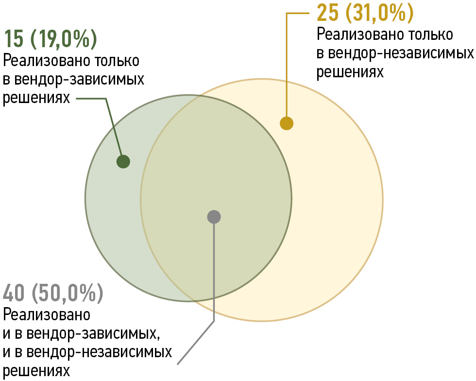
На рис. 1 представлено распределение функций между вендор-зависимыми и вендор-независимыми решениями; около половины из них реализовано в обоих типах ПО.

Рис. 1. Количество функций в зависимости от их реализации в вендор-зависимых и вендор-независимых решениях.
Практическая значимость основных функций вендор-зависимых решений
Установлено, что среди 17 проанализированных функций вендор-зависимых решений 6 не имели аналогов среди вендор-независимых (рис. 2).

Рис. 2. Практическая значимость основных функций вендор-зависимого программного обеспечения, по данным опроса врачей-рентгенологов: результаты представлены в виде процента общего числа опрошенных.
Следует отметить, что шесть функций вендор-зависимого ПО, не имеющих аналогов в вендор-независимых решениях (35,3%), используют менее 40% врачейрентгенологов. Наибольшую практическую значимость среди них имеют функции анализа сосудов, а также оценки перфузии мозга, тогда как наименьшую — функция выявления рака толстой кишки.
Из одиннадцати функций вендор-зависимого ПО, для которых предусмотрены аналоги в вендорнезависимом ПО (64,7%), шесть востребованы более чем у 50% врачей-рентгенологов. Наименее значимой в этой группе была функция оценки нарушений воздушности лёгочной ткани.
ОБСУЖДЕНИЕ
У вендор-зависимого и вендор-независимого ПО имеются некоторые одинаковые функции, однако в обеих группах решений есть и свои уникальные.
Сравнительная оценка функционала вендор-зависимых и вендор-независимых решений
Как упоминалось выше, вендор-зависимые и вендорнезависимые решения для модальности «Рентгенография» обладают почти одинаковым набором функций. Среди них: обнаружение новообразований, инфекционно-воспалительных и травматических изменений и др.
Все решения для модальности «Маммография» сосредоточены на выявлении признаков новообразований молочных желёз.
Наибольшие отличия в отношении функциональности характерны для вендор-зависимых и вендор-независимых решений для модальностей «Компьютерная томография» и «Магнитно-резонансная томография».
Следует отметить, что для компьютерной томографии органов грудной клетки среди вендор-зависимого ПО мало функций по обнаружению заболеваний лёгких (отсутствует детекция туберкулёза, саркоидоза, бронхоэктатической болезни и др.). При этом в вендорзависимом ПО имеются уникальные функции для анализа сердечно-сосудистой системы: анализ и 3D-моделирование сосудов, автоматическая детекция тромбов, спектральный анализ сосудов и сердца и др. Обратная картина характерна для вендор-независимого ПО.
Для компьютерной томографии органов брюшной полости вендор-зависимые решения предлагают функции по выявлению полипов и рака толстой кишки. В то же время вендор-независимые решения такими возможностями не располагают, однако обеспечивают выявление признаков мочекаменной болезни, а также новообразований надпочечников и почек.
При магнитно-резонансной томографии головного мозга вендор-зависимые решения позволяют оценивать перфузию, но не способны выявлять рассеянный склероз и новообразования, которые могут быть обнаружены с помощью вендор-независимого ПО.
Кроме того, вендор-независимое ПО обладает уникальными функциями выявления очаговых изменений костной структуры позвонков, а также протрузий, грыж межпозвонковых дисков и стеноза позвоночного канала.
Таким образом, наибольшие различия в части функционала между вендор-зависимым и вендор-независимым ПО наблюдают для модальностей «Компьютерная томография» и «Магнитно-резонансная томография».
Сравнительная оценка преимуществ и недостатков вендор-зависимых и вендор-независимых решений
Вендор-зависимое ПО поставляет производитель диагностического оборудования и рабочих станций. С этим связаны его определённые преимущества:
- полная совместимость всех компонентов;
- сервисная поддержка;
- отсутствие необходимости содержать большой штат специалистов в области поддержки систем информационных технологий;
- наличие уникальных, востребованных у врачейрентгенологов функций, которые отсутствуют в вендор-независимом ПО.
К недостаткам вендор-зависимого ПО можно отнести:
- высокую стоимость;
- невозможность его установки на рабочие станции от другого производителя;
- ограниченность функционала отдельных модулей.
По данным литературы, отмечают высокую частоту ложноположительных срабатываний такого ПО и его неравномерную эффективность в клинической практике [7–12]. Это может быть обусловлено тем, что, в отличие большинства вендор-независимых решений, основанных на методах глубокого обучения, многие вендор-зависимые решения опираются на признаки, выделенные экспертами вручную или полуавтоматически, и, следовательно, не учитывают всего многообразия [13, 14]. Однако стоить отметить, что некоторые вендор-зависимые решения всё же обладают высокой диагностической точностью. В последние годы появляются и вендор-зависимые решения, основанные на глубоком обучении, которые по точности не уступают вендор-независимому ПО. Тем не менее на данный момент их регулярная оценка качества проводится не всегда. Ещё одним недостатком вендорзависимого ПО может быть отсутствие комплексных решений, позволяющих выявлять основные виды патологических изменений при использовании определённой модальности и анатомической области (например, по результатам компьютерной томографии органов грудной клетки, брюшной полости и др.).
Преимуществами вендор-независимых решений являются регулярные улучшения их качества, которые быстро становятся доступны конечным пользователям, а также наличие большого количества уникальных функций. Среди вендор-независимого ПО появляется всё больше комплексных решений, которые способны обнаруживать все основные виды патологии по данным определённой модальности и анатомической области. Ещё одним преимуществом вендорнезависимых решений является гибкая система оплаты, которая подразумевает, что стоимость использования зависит от числа обработанных исследований18.
Необходимость дополнительной инфраструктуры можно отнести к недостаткам вендор-независимых решений. Например, небольшие медицинские организации без стабильных высокоскоростных каналов связи не смогут воспользоваться «облачными» решениями. Локальная установка некоторых вендор-независимых решений теоретически возможна, однако на практике качество их работы может быть значительно ниже, чем заявлено разработчиками [15–18]. Следует отметить, что для решения этой проблемы в России с 2020 года активно создают инфраструктуру, которая позволяет обеспечить «бесшовное» внедрение этих решений в лучевую диагностику всех регионов, а научно-обоснованная система технологических и клинических мониторингов позволяет гарантировать их высокое и стабильное качество работы19 [19–22].
Помимо всего вышеперечисленного, стоит упомянуть и о перспективах развития вендор-зависимого и вендор-независимого ПО. Многие вендор-зависимые решения основаны на алгоритмах, опирающихся на ограниченный набор признаков, выбранных экспертами вручную или полуавтоматически. Для увеличения метрик их диагностической точности необходима доработка или изменение алгоритмов, лежащих в их основе, что представляется очень трудоёмким. В связи с вышеизложенным, в последнее время при разработке как вендор-зависимых, так и вендор-независимых решений компании используют глубокое обучение, поскольку оно позволяет относительно быстро повысить метрики диагностической точности за счёт дообучения моделей на новых наборах данных [23–25]. Однако следует иметь в виду, что для обеспечения быстрого дообучения необходимы соответствующие вычислительные мощности.
Вендор-зависимые и вендор-независимые решения: основные подходы к выбору
Очевидна необходимость наличия системы поддержки принятия врачебных решений на АРМ врача-рентгенолога, поскольку она позволяют повысить качество диагностики и уменьшить время описания исследований [21, 26].
По нашему мнению, выбор решения в значительной степени будет обусловлен типом медицинской организации, в которой планируется применять данное ПО. При осуществлении выбора мы бы рекомендовали должностным лицам в сфере организации здравоохранения (министрам, главным врачам, их заместителям и др.) учитывать следующие типы медицинских организаций.
Амбулаторно-поликлинические медицинские организации
Одна из основных функций таких организаций — профилактика заболеваний. Соответственно в них выполняют большое количество профилактических скрининговых исследований — флюорографических/рентгенографических и маммографических. Как было показано выше, вендор-зависимое и вендор-независимое ПО для этих модальностей обладают примерно одинаковым набором функций, поэтому критерием выбора станет стоимость использования в пересчёте на одно исследование.
Кроме того, в организациях амбулаторнополиклинического звена могут выполнять компьютерную и магнитно-резонансную томографию. С учётом того, что их проводят для выявления широкого спектра заболеваний органов грудной клетки, брюшной полости, позвоночника и других областей, в данной ситуации предпочтительнее использование вендор-независимого ПО, поскольку именно оно обладает соответствующими функциями.
Референс-центры (дистанционные консультативные центры лучевой диагностики)
Согласно Приказу Министерства Здравоохранения Российской Федерации № 560н от 9 июня 2020 г. «Об утверждении Правил проведения рентгенологических исследований»20, к функциям референс-центров относят:
- анализ результатов рентгенологических исследований;
- организация и проведение двойного просмотра результатов массовых профилактических осмотров (скрининга), в том числе с использованием автоматизированных систем;
- выявление и анализ причин расхождений в результатах рентгенологических исследований с разработкой и реализацией мероприятий по обеспечению качества и т. д.
В связи с этим подходы к выбору систем поддержки принятия врачебных решений могли бы быть во многом сходны с теми, что описаны для амбулаторнополиклинических медицинских организаций. Вместе с тем необходимо учитывать особенности организации работы каждого конкретного референс-центра.
Так, в Москве лучевые исследования выполняют в амбулаторно-поликлинических медицинских организациях, а затем результаты попадают в единый радиологический информационный сервис. Именно в нём врачирентгенологи московского референс-центра осуществляют просмотр и описание исследований. При такой схеме организации лучевой диагностики использование вендор-зависимого ПО становится невозможно, поскольку АРМ врачей не связаны напрямую с диагностическим оборудованием.
Это не позволяет врачам использовать уникальные функции вендор-зависимого ПО, но как показал опрос, они обладают той или иной степенью практической значимости менее чем для 40% врачей.
Стационарные медицинские организации
Как правило, такие медицинские организации специализируются на оказании медицинской помощи пациентам по одному или нескольким профилям. В связи с этим при отсутствии альтернативы предлагается выбирать те решения, которые обладают необходимыми функциями. Например, в сердечно-сосудистом центре целесообразно использовать вендор-зависимое ПО, ориентированное на анализ сердечно-сосудистой системы. В учреждениях, специализирующихся на дегенеративных заболеваниях позвоночника, более оправдано применение вендорнезависимого ПО с соответствующими функциями.
В случаях, когда необходимые инструменты представлены в обоих типах решений, выбор определяется уровнем развития инфраструктуры и принципом экономической целесообразности.
Таким образом, оба подхода к применению ПО для анализа лучевых исследований — вендорзависимый или вендор-независимый — остаются актуальными, а предпочтение конкретного варианта определяется особенностями медицинской организации.
Ограничения работы
Представленный анализ проводили на примере системы здравоохранения Москвы. Выбор предлагаемых подходов к использованию ПО для анализа лучевых исследований в других субъектах Российской Федерации и странах рекомендуется осуществлять с учётом особенностей местных систем здравоохранения.
При опросе врачи-рентгенологи оценивали значимость функций только тех решений, которые доступны им по месту работы в конкретной медицинской организации. Авторский опросник не проходил процедуру валидности и надёжности, что могло повлиять на точность измерений конструкта «практическая значимость».
Кроме того, исследование проводили в период с сентября 2023 г. по март 2024 г., в свою очередь, рынок рассматриваемых продуктов быстро меняется, а разработчики добавляют новые функции. Именно поэтому перед принятием решений о закупке того или иного ПО рекомендуем проводить дополнительный анализ рынка рассматриваемых решений.
Перечень наименований функций ПО сформулирован в соответствии с базовыми диагностическими требованиями [27], а также информацией, представленной на сайтах компаний-производителей.
ЗАКЛЮЧЕНИЕ
Таким образом, только половина функций встроенного вендор-зависимого и вендор-независимого ПО, доступных для использования в Москве, совпадают.
Уникальные функции встроенного вендор-зависимого и вендор-независимого ПО определяют выбор в пользу определённого решения в зависимости от потребностей врачей-рентгенологов конкретной медицинской организации. В случае необходимости возможно одновременно использовать оба типа ПО. Почти 2/3 функций встроенного вендор-зависимого ПО (64,7%), используемого врачамирентгенологами Москвы, могут быть реализованы с помощью вендор-независимых решений. В таких условиях выбор ПО следует осуществлять с учётом развитости инфраструктуры и экономической целесообразности.
ДОПОЛНИТЕЛЬНАЯ ИНФОРМАЦИЯ
Приложение 1. Опросник для оценки практической значимости основных функций вендор-зависимого программного обеспечения. doi: 10.17816/DD631519-4379059
Приложение 2. Функциональные возможности встроенных вендор-зависимых и вендор-независимых решений. doi: 10.17816/DD631519-4379066
Вклад авторов. Ю.А. Васильев — концепции работы; Е.А. Славущева — сбор и анализ литературных данных, написание и редактирование текста рукописи; М.А. Зеленова — сбор и анализ литературных данных, создание опросника и проведение опроса; И.М. Шулькин, К.М. Арзамасов — концепция работы, редактирование текста рукописи. Все авторы одобрили рукопись (версию для публикации), а также согласились нести ответственность за все аспекты работы, гарантируя надлежащее рассмотрение и решение вопросов, связанных с точностью и добросовестностью любой её части.
Благодарности. Авторы благодарят Р.Н. Ахметова за консультацию по текущим потребностям врачей в использовании систем поддержки принятия врачебных решений, а также С.В. Михайлина и Д.В. Козлова за аналитику рынка автоматизированных рабочих мест врачейрентгенологов.
Этическая экспертиза. Данное исследование основано на результатах Эксперимента по использованию инновационных технологий в области компьютерного зрения для анализа медицинских изображений и дальнейшего применения в системе здравоохранения города Москвы, утверждённого этическим комитетом (выписка из протокола № 2 НЭК МРО РОРР от 20.02.2020), также зарегистрированного на ClinicalTrials (NCT04489992).
Источник финансирования. Данная статья подготовлена авторским коллективом в рамках научно-исследовательской и опытноконструкторской работы «Разработка платформы повышения качества ИИ-Сервисов для медицинской диагностики» (ЕГИСУ: № 123031400006-0) в соответствии с Приказом № 1196 от 21 декабря 2022 г. «Об утверждении государственных заданий, финансовое обеспечение которых осуществляется за счёт средств бюджета города Москвы государственным бюджетным (автономным) учреждениям подведомственным Департаменту здравоохранения города Москвы, на 2023 год и плановый период 2024 и 2025 годов» Департамента здравоохранения города Москвы.
Раскрытие интересов. Авторы заявляют об отсутствии отношений, деятельности и интересов за последние три года, связанных с третьими лицами (коммерческими и некоммерческими), интересы которых могут быть затронуты содержанием статьи.
Оригинальность. При создании настоящей работы авторы не использовали ранее опубликованные сведения (текст, иллюстрации, данные).
Доступ к данным. Все данные, полученные в настоящем исследовании, доступны в статье и в приложении к ней. В частности, в Приложении 1 и 2.
Генеративный искусственный интеллект. При создании настоящей статьи технологии генеративного искусственного интеллекта не использовали.
Рассмотрение и рецензирование. Настоящая работа подана в журнал в инициативном порядке и рассмотрена по обычной процедуре. В рецензировании участвовали два члена редакционной коллегии журнала.
ADDITIONAL INFORMATION
Supplement 1: Questionnaire for assessing the practical significance of the main functions of vendor-dependent software. doi: 10.17816/DD631519-4379064
Supplement 2: Functionality of embedded vendor-dependent and vendor-independent solutions. doi: 10.17816/DD631519-4379067
Author contributions: Yu.A. Vasilev: conceptualization; E.A. Slavushcheva: data curation, writing—original draft, review & editing; M.A. Zelenova: data curation, survey creation and implementation; I.M. Shulkin, K.M. Arzamasov: conceptualization, writing—review & editing. All the authors approved the final version of the manuscript for publication and agreed to be accountable for all aspects of the work, ensuring that questions related to the accuracy or integrity of any part of the work are appropriately investigated and resolved.
Acknowledgments: The authors would like to thank R.N. Akhmetov for consultation on the current demands of healthcare providers regarding clinical decision–support systems, and S.V. Mikhailin and D.V. Kozlov for analyzing the market for automated radiology workstations.
Ethics approval: This study was based on the data from the approved Experiment on the use of innovative computer vision technologies for analysis of medical images in the Moscow healthcare system (extract from Minutes No. 2 of the Independent Ethics Committee of the Moscow Regional Branch of the Russian Society of Radiographers and Radiologists dated February 20, 2020), registered at ClinicalTrials (NCT04489992).
Funding sources: This article was part of the research and development project “Development of a platform for improving the quality of AI services for medical diagnostics” (EGISU No. 123031400006-0) under Moscow City Health Department Order No. 1196 On the Approval of State Assignments Funded from the Budget of the City of Moscow for State Budgetary (Autonomous) Institutions Subordinate to the Moscow City Health Department for 2023 and the Planned Period of 2024–2025, dated December 21, 2022.
Disclosure of interests: The authors have no relationships, activities, or interests for the last three years related to for-profit or not-for-profit third parties whose interests may be affected by the content of the article.
Statement of originality: No previously published material (text, images, or data) was used in this study or article.
Data availability statement: All data obtained in this study are available in the article and its supplementary material. In particular, in Supplements 1 and 2.
Generative AI: No generative artificial intelligence technologies were used to prepare this article.
Provenance and peer-review: This article was submitted unsolicited and reviewed following the standard procedure. The peer review process involved two members of the Editorial Board.
1 Здесь и далее по тексту вендор-зависимое ПО — решения, которые предлагают поставщики диагностического оборудования (рентгеновских, маммографических аппаратов, компьютерных и магнитно-резонансных томографов), тогда как вендор-независимое ПО — решения, предлагаемые сторонними компаниями, то есть теми, кто диагностическое оборудование не производит.
2 Каталог ИИ сервисов [интернет]. В: Центр диагностики и телемедицины. Режим доступа: https://mosmed.ai/service_catalog/ Дата обращения: 24.10.2024.
3 Программное обеспечение для медицинской визуализации: каталог [интернет]. Medical Expo. Режим доступа: https://www.medicalexpo.ru/cat/radiologia/ Дата обращения: 01.08.2023.
4 CAD4TB: AI-Powered Tuberculosis Detection Software [интернет]. Режим доступа: https://delft.care/cad4tb/ Дата обращения: 01.08.2024.
5 Philips IntelliSpace Portal 12 [интернет]. Режим доступа: https://www.philips.com.om/healthcare/product/ Дата обращения: 30.10.2024.
6 Gehealthcare [интернет]. Режим доступа: https://www.gehealthcare.com/products/healthcare-it Дата обращения: 30.10.2024.
7 Siemens-healthineers [интернет]. Режим доступа: https://www.siemens-healthineers.com/digital-health-solutions/ Дата обращения: 30.10.2024.
8 Canon [интернет]. Режим доступа: https://us.medical.canon/specialties/medical-oncology-solutions/ Дата обращения: 30.10.2024.
9 Hologic [интернет]. Режим доступа: https://www.hologic.com/hologic-products/breast-health-solutions/ Дата обращения: 30.10.2024.
10 Aidoc [интернет]. Режим доступа: https://www.aidoc.com/solutions/#aipowered/ Дата обращения: 30.10.2024.
11 RADinfo SYSTEMS [интернет]. Режим доступа: https://radinfosystems.com/products.html Дата обращения: 30.10.2024.
12 Sectra Medical [интернет]. Режим доступа: https://medical.sectra.com/product/sectra-radiology-pacs-ris/ Дата обращения: 30.10.2024.
13 Visage Imaging [интернет]. Режим доступа: https://visageimaging.com/ Дата обращения: 30.10.2024.
14 TeraRecon [интернет]. Режим доступа: https://www.terarecon.com/ Дата обращения: 30.10.2024.
15 Agfa HealthCare [интернет]. Режим доступа: https://www.agfa.com/he/russia/ru/internet/main/ Дата обращения: 30.10.2024.
16 Synapse [интернет]. Режим доступа: https://synapse-emea.fujifilm.com/discovering-synapse-workflow.html Дата обращения: 30.10.2024.
17 Merge PACS — American Medical Imaging [интернет] Режим доступа: https://ami-ii.com/products/merge-pacs/ Дата обращения: 30.10.2024.
18 Приказ Департамента Здравоохранения Москвы № 134 от 24 февраля 2022 г. «Об утверждении Порядка и условий проведения эксперимента по использованию инновационных технологий в области компьютерного зрения для анализа медицинских изображений и дальнейшего применения в системе здравоохранения города Москвы». Режим доступа: https://mosmed.ai/ai/docs/ Дата обращения: 10.02.2024.
19 Указ Президента Российской Федерации № 490 от 10 октября 2019 г. «О развитии искусственного интеллекта в Российской Федерации» (с изменениями и дополнениями). Режим доступа: https://base.garant.ru/72838946/ Дата обращения: 10.02.2024.
20 Приказ Министерства здравоохранения Российской Федерации № 560н от 9 июня 2020 г. «Об утверждении Правил проведения рентгенологических исследований». Режим доступа: https://base.garant.ru/74632238/ Дата обращения: 10.02.2024.
About the authors
Yuriy A. Vasilev
Research and Practical Clinical Center for Diagnostics and Telemedicine Technologies; National Medical and Surgical Center named after N.I. Pirogov
Email: VasilevYA1@zdrav.mos.ru
ORCID iD: 0000-0002-5283-5961
SPIN-code: 4458-5608
MD, Cand. Sci. (Medicine)
Russian Federation, Moscow; MoscowEkaterina A. Slavushcheva
Research and Practical Clinical Center for Diagnostics and Telemedicine Technologies
Email: SlavuschevaEA1@zdrav.mos.ru
ORCID iD: 0009-0009-1315-0829
MD
Russian Federation, MoscowMaria A. Zelenova
Research and Practical Clinical Center for Diagnostics and Telemedicine Technologies
Email: ZelenovaMA@zdrav.mos.ru
ORCID iD: 0000-0001-7458-5396
SPIN-code: 3823-6872
Cand. Sci. (Biology)
Russian Federation, MoscowIgor M. Shulkin
Research and Practical Clinical Center for Diagnostics and Telemedicine Technologies
Email: ShulkinIM@zdrav.mos.ru
ORCID iD: 0000-0002-7613-5273
SPIN-code: 5266-0618
MD, Cand. Sci. (Medicine)
Russian Federation, MoscowKirill M. Arzamasov
Research and Practical Clinical Center for Diagnostics and Telemedicine Technologies; MIREA — Russian Technological University
Author for correspondence.
Email: ArzamasovKM@zdrav.mos.ru
ORCID iD: 0000-0001-7786-0349
SPIN-code: 3160-8062
MD, Dr. Sci. (Medicine)
Russian Federation, Moscow; MoscowReferences
- Shulkin IM, Vladzimirsky AV. Data-based Management in Imaging: Evaluation of the Performance of a Unified Radiological Information Service Model. Manager Zdravoochranenia. 2022;(7):68–80. doi: 10.21045/1811-0185-2022-7-68-80 EDN: IYPKHK
- Finakov AS. Information Technologies in Medicine. Scientific Journal of Academy. 2022;(3):63–67. EDN: WSNKRW
- Morozov SP, Vladzymyrskyy AV, Ledikhova NV, et al. Moscow Experiment on Computer Vision in Radiology: Involvement and Participation of Radiologists. Medical Doctor and IT. 2020;(4):14–23. doi: 10.37690/1811-0193-2020-4-14-23 EDN: VEWGXO
- Guo Z, Xie J, Wan Y, et al. A Review of the Current State of the Computer-Aided Diagnosis (CAD) Systems for Breast Cancer Diagnosis. Open Life Sciences. 2022;17(1):1600–1611. doi: 10.1515/biol-2022-0517 EDN: RSGRVD
- Fujita H. AI-based Computer-Aided Diagnosis (AI-CAD): The Latest Review to Read First. Radiological Physics and Technology. 2020;13(1):6–19. doi: 10.1007/s12194-019-00552-4 EDN: JHXXPS
- Mayo RC, Kent D, Sen LC, et al. Reduction of False-Positive Markings on Mammograms: a Retrospective Comparison Study Using an Artificial Intelligence-Based CAD. Journal of Digital Imaging. 2019;32(4):618–624. doi: 10.1007/s10278-018-0168-6 EDN: BHBLVP
- Kohli A, Jha S. Why CAD Failed in Mammography. Journal of the American College of Radiology. 2018;15(3):535–537. doi: 10.1016/j.jacr.2017.12.029
- Lehman CD, Wellman RD, Buist DSM, et al. Diagnostic Accuracy of Digital Screening Mammography With and Without Computer-Aided Detection. JAMA Internal Medicine. 2015;175(11):1828. doi: 10.1001/jamainternmed.2015.5231
- Fenton JJ, Taplin SH, Carney PA, et al. Influence of Computer-Aided Detection on Performance of Screening Mammography. New England Journal of Medicine. 2007;356(14):1399–1409. doi: 10.1056/NEJMoa066099
- Gilbert FJ, Astley SM, Gillan MGC, et al. Single Reading with Computer-Aided Detection for Screening Mammography. New England Journal of Medicine. 2008;359(16):1675–1684. doi: 10.1056/NEJMoa0803545
- Tchou PM, Haygood TM, Atkinson EN, et al. Interpretation Time of Computer-aided Detection at Screening Mammography. Radiology. 2010;257(1):40–46. doi: 10.1148/radiol.10092170
- Philpotts LE. Can Computer-aided Detection Be Detrimental to Mammographic Interpretation? Radiology. 2009;253(1):17–22. doi: 10.1148/radiol.2531090689
- Khanna NN, Maindarkar MA, Viswanathan V, et al. Economics of Artificial Intelligence in Healthcare: Diagnosis vs. Treatment. Healthcare. 2022;10(12):2493. doi: 10.3390/healthcare10122493 EDN: UDQHMZ
- Xing X, Zhao X, Wei H, Li Y. Diagnostic Accuracy of Different Computer-Aided Diagnostic Systems for Prostate Cancer Based on Magnetic Resonance Imaging. Medicine. 2021;100(3):e23817. doi: 10.1097/md.0000000000023817 EDN: EUAPKQ
- Hadjiiski L, Cha K, Chan HP, et al. AAPM Task Group Report 273: Recommendations on Best Practices for AI and Machine Learning for Computer-Aided Diagnosis in Medical Imaging. Medical Physics. 2023;50(2):e1–e24. doi: 10.1002/mp.16188 EDN: RFUOFA
- Roberts M, Driggs D, Thorpe M, et al; AIX-COVNET. Common Pitfalls and Recommendations for Using Machine Learning to Detect and Prognosticate for COVID-19 Using Chest Radiographs and CT Scans. Nature Machine Intelligence. 2021;3(3):199–217. doi: 10.1038/s42256-021-00307-0 EDN: YFUJIA
- Gu Y, Chi J, Liu J, et al. A Survey of Computer-Aided Diagnosis of Lung Nodules from CT Scans Using Deep Learning. Computers in Biology and Medicine. 2021;137:104806. doi: 10.1016/j.compbiomed.2021.104806 EDN: LPGPQM
- Aggarwal R, Sounderajah V, Martin G, et al. Diagnostic Accuracy of Deep Learning in Medical Imaging: A Systematic Review and Meta-analysis. NPJ Digital Medicine. 2021;4(1):65–65. doi: 10.1038/s41746-021-00438-z EDN: RYOQDM
- Vladzimirskyy AV, Vasilev YuA, Arzamasov KM, et al. Computer Vision in Radiation Diagnostics: The First Stage of the Moscow Experiment. Moscow: Izdatel'skie resheniya; 2023. ISBN: 978-5-0059-3043-9 (In Russ.) EDN: FOYLXK
- Morozov SP, Vladzymyrskyy AV, Shulkin IM, et al. Feasibility of Using Artificial Intelligence in Radiology (First Year of Moscow Experiment on Computer Vision). Medical Doctor and IT. 2022;(1):12–29. doi: 10.25881/18110193_2022_1_12 EDN: UCHWWP
- Vasilev YuA, Vladzymyrskyy AV, Bondarchuk DV, et al. Importance of Artificial Intelligence Technologies to Prevent Defects in Radiologist’s Practice. Medical Doctor and IT. 2023;(2):16–27. doi: 10.25881/18110193_2023_2_16 EDN: SYZAOQ
- Vasilev YuA, Vladzimirskyy AV, Omelyanskaya OV, et al. Review of Meta-analyses on the Use of Artificial Intelligence in Radiology. Medical Visualization. 2024;28(3):22–41. doi: 10.24835/1607-0763-1425 EDN: QYASNZ
- Kim DW, Jang HY, Kim KW, et al. Design Characteristics of Studies Reporting the Performance of Artificial Intelligence Algorithms for Diagnostic Analysis of Medical Images: Results from Recently Published Papers. Korean Journal of Radiology. 2019;20(3):405–410. doi: 10.3348/kjr.2019.0025
- Nam JG, Hwang EJ, Kim J, et al. AI Improves Nodule Detection on Chest Radiographs in a Health Screening Population: A Randomized Controlled Trial. Radiology. 2023;307(2):e221894. doi: 10.1148/radiol.221894 EDN: ZXSHYF
- Yu X, Wang J, Hong QQ, et al. Transfer Learning for Medical Images Analyses: A survey. Neurocomputing. 2022;489:230–254. doi: 10.1016/j.neucom.2021.08.159 EDN: NQEUQB
- Vladzymyrskyy AV, Kudryavtsev ND, Kozhikhina DD, et al. Effectiveness of Using Artificial Intelligence Technologies for Dual Descriptions of the Results of Preventive Lung Examinations. Russian Journal of Preventive Medicine. 2022;25(7):7–15. doi: 10.17116/profmed2022250717 EDN: JNUMFN
- Morozov SP, Abuladze LR, Andreychenko AE, et al. Basic Recommendations for the Operation of Artificial Intelligence Services for Radiation Diagnostics. Moscow: Research and Practical Clinical Center for Diagnostics and Telemedicine Technologies; 2022. (In Russ.) EDN: EHQKPW
Supplementary files

