

")


卷 2, 编号 1 (2021)
- 年: 2021
- ##issue.datePublished##: 30.04.2021
- 文章: 8
- URL: https://jdigitaldiagnostics.com/DD/issue/view/3394
- DOI: https://doi.org/10.17816/DD.21

原创性科研成果
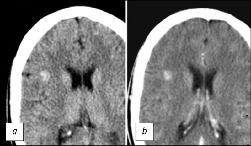
CT诊断的准确率,以确定COVID-19患者的住院需求
摘要
论证:在俄罗斯联邦,为了检测COVID-19肺炎及其并发症和与其他肺部疾病的鉴别诊断,以及对患者进行分类,使用了胸部CT,并在CT 0–4的半定量视觉尺度上评估变化。尽管胸部CT广泛使用,但其用于确定COVID-19患者住院需求的诊断准确性的数字指标目前尚不清楚。
目的: 是确定该量表的敏感性、特异性、阳性预测值、阴性预测值。
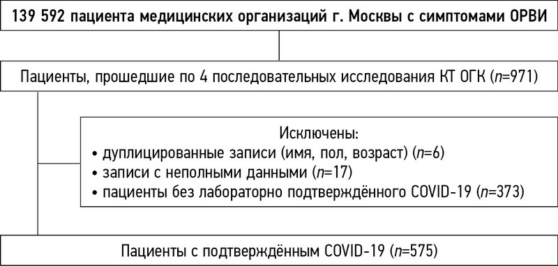
材料与方法:研究涉及575名经实验室确诊的COVID-19患者(55%为女性),年龄为57.2±13.9岁。对于每个患者,进行了4次连续的胸部CT研究,并对疾病的严重程度进行了CT评分(0–4)。根据既往CT研究结果,将敏感性和特异性作为患者病情恶化或改善的条件概率进行计算。为计算阳性预测值(PPV)和阴性预测值(NPV),对COVID-19在莫斯科的流行情况进行了估计。2020年3月6日至11月28日期间所有COVID-19病例的数据来自俄国国家管理的保护消费者服务机构(Rospotrebnadzor)网站。使用了许多具有不同参数的ARIMA和EST模型来选择与现有数据最匹配的模型,并预测发病率的发展。
结果:0–4 CT分级的中位特异性为69%,敏感性为92%。描述莫斯科流行病学情况的最佳统计模型是ARIMA(0,2,1)。经计算,预测年发病率为9.6%,PPV值为56,NPV值为97%。
结果:Yuden指数最大的阶段出现在胸部CT第一次研究和第二次研究之间,此时样本中大多数患者表现出临床病情恶化的趋势。0–4 CT分级可以安全地排除轻、中度病程(CT0、CT1类)患者的病理变化发展,有助于优化患者在疫情不利的情况下住院。
 5-16
5-16


学生对介入放射学硕士课程电子学习的看法:一项学员调查
摘要
目的:探讨介入放射学硕士研究生对远程教育的看法。
方法:硕士的核心课程分为3个电子学习模块和2个电子学习+实践培训模块。电子学习通过一个在线会议平台开展,该平台可实现同步培训,提供实时授课,并在专门的网站上录制播放。提供实地操作培训,可帮助介入放射科医生对患者执行介入手术治疗。目前已准备了包括12个问题的在线调查,用以确定培训质量。学生通过5分制量表说明其对电子学习和实践培训影响的认同程度,并计算认同程度的平均分数。
结果:本系列研究有16名学员参加。62.5%的学员在公立非学术性医院工作,80%的学员已经以主刀身份执行超过300次介入手术。
学员一致认为,电子学习模块的主要优势是能够方便讲座出勤(68.8%),其次是培训成本低(18.8%)。没有学生对陈述的评分低于3分。81.3%的学员一致认为,该硕士课程达到了学习预期。
讨论:学员非常满意,并愿意向其他同事推荐该硕士课程。该复合型硕士课程教育获高度称赞,并且可能成为未来介入放射学(IR)可以采用的模式。
17-26
人工智能如何影响胸部CT扫描对COVID-19中肺损伤的评估?
摘要
理由:在大流行期间,计算机断层扫描(CT)是评估与COVID-19相关的肺部变化的主要工具之一。莫斯科的放射学家使用了经过调整的KT0-4量表,根据计算机断层扫描技术,通过视觉评估了一般病情严重程度对COVID-19中肺部改变的放射学征象的性质和严重程度的依赖性。大量的研究中,医生可能会遗漏发现结果并在评估肺损伤量方面犯错误,因此在大流行期间,在门诊医疗中使用AI服务可能很有用。
目的:比较放射科医生形成的CT0-4类别的分布与AI服务处理的结果以及没有AI服务形成的类别的比较。方法:回顾性研究,ClinicalTrials.gov(NCT04489992)。DZM的门诊医疗组织中,分析了从CT0-4类别进行的一次CT扫描的结果,分析时间为:2020年4月8日至2020年1月12日,以及11月(2020年11月1日至2020年1月12日)。根据标准协议在48台计算机断层扫描仪上执行CT,并通过ERIS处理。测试组包括由AI服务处理的CT,对照组为不包含AI的CT。分析包括5种AI服务:RADlogics COVID-19(美国RADLogics),COVID-IRA(俄罗斯的IRA实验室),Care Mentor AI,COVID(俄罗斯的CareMentor AI),第三意见。CT-COVID-19英寸(第三意见,俄罗斯),COVID-MULTIVOX(俄罗斯伽马迈德)。AI服务是随机编码的。
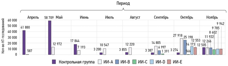
结果:分析了260594例患者的CT扫描结果(m / f%= 44/56,平均年龄-49.5)。测试组包括115,618次CT扫描,对照组-144976。根据特定的AI服务,对于 CT-0类别的不同子组,其设置比对照组少2.3%至18.5%。与未使用AI相比,将CT3-4类别设置为比不使用AI少4.7%至27.6%,并且将CT-4类别与不使用AI设置成从40%至60%(p <0.0001)。
对于11月(从01.11.2020到01.12.2020),分析了41386名患者的CT扫描结果(m / f%= 44/56,平均年龄-53.2岁)。测试组包括28881 CT扫描,对照组-12505。根据特定的AI服务,对于CT-0类别的不同子组,其设置比对照组小1%至2.6%。显示的CT3-4类别比没有使用AI的类别多出0.2%至15.7%; 类别CT-4设置为比不使用AI时少25%(p = 0.001)。
结论:在门诊基础上将AI服务用于主要CT扫描会导致CT-0和CT3-4数量减少,从而影响管理COVID-19患者的策略。
27-38
系统评价
脑洞畸形的放射诊断
摘要
目前,脑海绵状畸形是相当普遍的血管病理:近年来发现的病例数量急剧增加。这是由于将其引入临床实践并广泛传播了现代神经成像方法,例如计算机断层扫描(CT)和磁共振成像(MRI)断层扫描。CT和MRI出现之前,很难诊断出这种病理,诊断通常是在术中或根据尸检数据进行的。文献综述致力于脑海绵状畸形(CM)的放射学诊断。分析了神经影像学方法对海绵状畸形的诊断的重要性,以及使用MRI对骨髓进行可视化的重要性。相比于这种病理学的其他神经影像学检查方法,MRI具有优势。根据形态学底物,对MRI的脉冲序列和各种类型灶的信号特征进行了表征。分析 SWI (susceptibility weighted imaging)序列的值 用于检测家族性CM病例中的多灶性病变。对MRI的主要脉冲序列进行可视化以研究海绵状畸形的研究将有助于优化协议算法,以便及时诊断这种病理状况并选择治疗策略。
39-48
技术说明
标准医疗日期(MosMedData)独立外部评价的算法在诊断的人工智能基础上
摘要
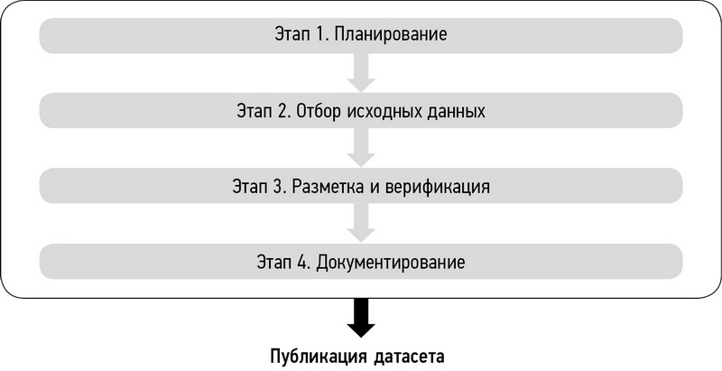
这篇文章介绍了一个独特的方法来创建附加说明的医疗日期,以测试基于人工智能技术的诊断解决方案。描述了数据集形成的四个阶段-计划,初始数据选择,标记和验证,文档。所举的例子是根据上述日期方法建立的。该方法是广泛而普遍的,因此可以应用于医学和卫生的其他领域,它是由人工智能技术和高数据技术的自动化和发展
49-66
临床病例及临床病例的系列
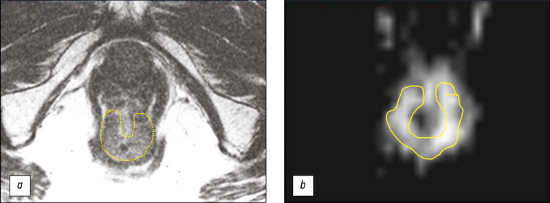
MRI评价1例直肠癌新辅助放化疗结果,辅以肿瘤T2WI结构分析(临床病例)
摘要
本文报告一名对新辅助化疗有良好反应的73岁下段壶腹直肠癌患者,采用积极动态随访策略(Watch & Wait策略)的临床病例。经过三年的定期随访,包括指状直肠检查、直肠镜检查和磁共振成像(MRI),表明肿瘤没有进展,得到了18F氟脱氧葡萄糖正电子发射断层摄影与计算机断层摄影的结果。结果显示直肠下段壶腹部有一个高代谢活动的部位(SUVmax 27.1),因此决定进行手术治疗。讨论手术范围时,考虑MRI资料,辅以T2WI分析结果,证实无疾病进展。患者接受了保留器官的经肛门肿瘤切除体积的治疗。手术准备的病理形态学检查确定了肠壁炎症改变和肿瘤的消失。本案例证明了标准调查体积在使用Watch & Wait策略时的有效性,以及使用T2WI分析来提高MRI评估肿瘤对放化疗反应的可靠性的可能性。
67-74
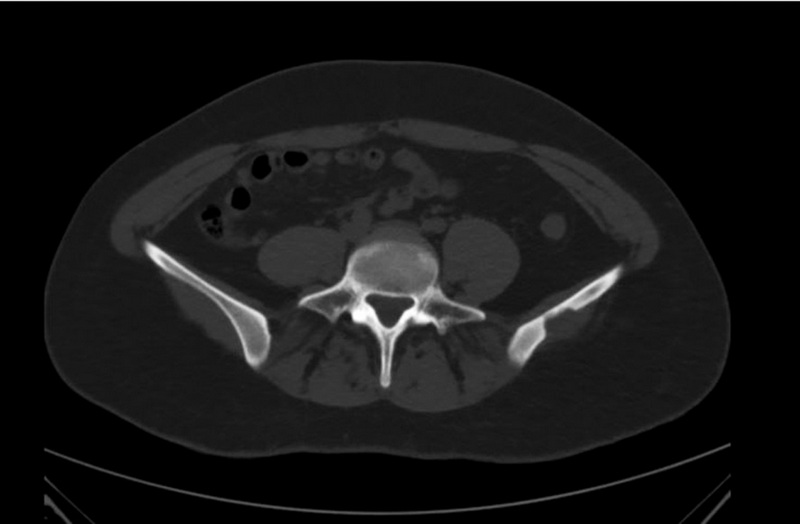
2例证实的孤立性嗜酸性肉芽肿 CT、MRI和18F-FDG PET/CT成像
摘要
本文介绍计算机诊断嗜酸性骨肉芽肿、磁共振和18F氟脱氧葡萄糖正电子发射断层扫描以及计算机断层扫描的两个临床观察。根据综合的放射学诊断研究和组织学证实,确诊为孤立性嗜酸性肉芽肿,两例患者都因怀疑原发性恶性骨肿瘤而入院。孤立性嗜酸性肉芽肿是一种相当罕见的疾病(不到1%的骨骼肿瘤体积形成病例)。最常见的是,嗜酸性肉芽肿见于头骨的顶骨和额骨,是一种溶骨体积的形成,逐渐增大。虽然大多数骨肿瘤可以通过X线摄影发现,计算机体层摄影术是首选,主要是因为它能很好地显示骨皮质层的破坏情况。计算机断层扫描和磁共振成像的诊断准确性可能不同。辐射和放射性核素诊断方法的复杂应用使能够缩小鉴别诊断的范围。在大多数病例中,现有的放射学诊断研究的特异性较低,不能做出准确的诊断,选择的方法仍然是活检后进行病理形态学检查。这些临床观察表明,当发现孤立的溶骨性病灶时,鉴别诊断需要包括嗜酸性肉芽肿。
75-82
致编辑的一封信
放射性核素诊疗一体化:一种新的个性化医学

摘要
放射性核素诊疗一体化允许使用各种放射性药物在体内进行分子成像(单光子发射计算机断层摄影术,正电子发射断层摄影术),并选择性地影响肿瘤引起的病理代谢过程的放射性核素治疗。自上世纪50年代以来,利用治疗诊断学的范式,在放射性碘的帮助下,成功地治疗了甲状腺毒症和甲状腺癌。近年来,由于核医学(在医疗机构回旋加速器、单光子发射计算机断层扫描/计算机断层扫描、正电子发射断层摄影术/计算机断层摄影术数量的增加)特别是放射药物的成功发展,放射性核素诊疗一体化在世界上发展非常迅速。基于177Lu、225Ac等放射性同位素的新型放射性配体的出现,刺激了大量(超过300)的前列腺癌、神经内分泌肿瘤、胰腺癌等恶性肿瘤的放射配体治疗的临床研究。基于靶向抗肿瘤药物的放射配体的开发是放射性核素诊疗一体化中最有前途的领域之一,这使得总结出一种放射药物的两种作用—信号级联抑制和辐射损伤。放射性核素诊疗一体化本质是多学科的、技术复杂的(同位素,放射性药物,单光子发射计算机断层扫描,正电子发射断层扫描)、整体的,并要求高能力和团队合作。放射性核素诊疗一体化和靶向放射药物的发展在俄罗斯尚处于起步阶段。主要的问题是这个领域缺乏专家:医生、物理学家、化学家、放射性药物学家、生物学家、遗传学家、工程师、程序员。医生和患者对放射性核素诊疗一体化可能性的认识不足也阻碍了其在俄罗斯临床应用的发展。
83-89
Регистрационный номер и дата принятия решения о регистрации СМИ: серия ПИ № ФС 77 - 79539 от 09 ноября 2020 г.