

")


Том 4, № 1 (2023)
- Жылы: 2023
- ##issue.datePublished##: 19.04.2023
- Мақалалар: 8
- URL: https://jdigitaldiagnostics.com/DD/issue/view/4561
- DOI: https://doi.org/10.17816/DD.41

Original Study Articles
Volumetry versus linear diameter lung nodule measurement: an ultra-low-dose computed tomography lung cancer screening study

Аннотация
BACKGROUND: The Dutch–Belgian Randomized Lung Cancer Screening Trial (NELSON) used a volume-based protocol and significantly reduced the prevalence of false-positive results (2.1%).
AIM: To compare the performance of manual linear diameter and semi-automated volumetric nodule measurement in the pilot project “Moscow Lung Cancer Screening” ultra-low-dose computed tomography pilot study.
MATERIALS AND METHODS: The study included individuals with a lung nodule of at least 4 mm on baseline-computed tomography of the Moscow lung cancer screening between February 2017 and February 2018, without verified lung cancer diagnosis until 2020. The radiation dose was selected individually and did not exceed 1 mSv. All scans were assessed by three blinded readers to measure the maximum and minimum transversal nodule diameter and extrapolated volume. As a reference value of size and volume, the average value from the results of expert measurements was obtained. A false-positive nodule was defined as a nodule <6 mm/<100 mm3 and a false-negative nodule as a nodule ≥6 mm/≥100 mm3.
RESULTS: Overall, 293 patients were included (166 men; mean age, 64.6 ± 5.3years); 199 lung nodules were <6 mm/<100 mm3 and 94 were ≥6 mm/≥100 mm3. Regarding volumetric measurements, 32 [10.9%; 4 false-positive, 28 false-negative], 29 [9.9%; 17 false-positive, 12 false-negative], and 30 [10.2%; 6 false-positive, 24 false-negative] nodule discrepancies were reported by readers 1, 2, and 3 respectively. For linear diameter measurement, 92 [65.5%; 107 false-positive, 85 false-negative], 146 [49.8%; 58 false-positive, 88 false-negative], and 102 [34.8%; 23 false-positive, 79 false-negative] nodule discrepancies were reported by readers 1, 2, and 3 respectively.
CONCLUSIONS: The use of lung nodule volumetry strongly reduces the number of false-positive and false-negative nodules compared with nodule diameter measurements, in an ultra-low-dose computed tomography lung cancer screening program.
 5-13
5-13


Technical Reports
Tele-ultrasound imaging using smartphones and single-board PCs
Аннотация
BACKGROUND: Mobile devices are widely available and their computational performance increases. Nonetheless, medicine should not be an exception: single-board computers and mobile phones are crucial aides in telehealth.
AIM: To explore tele-ultrasound scope using smartphones and single-board computers
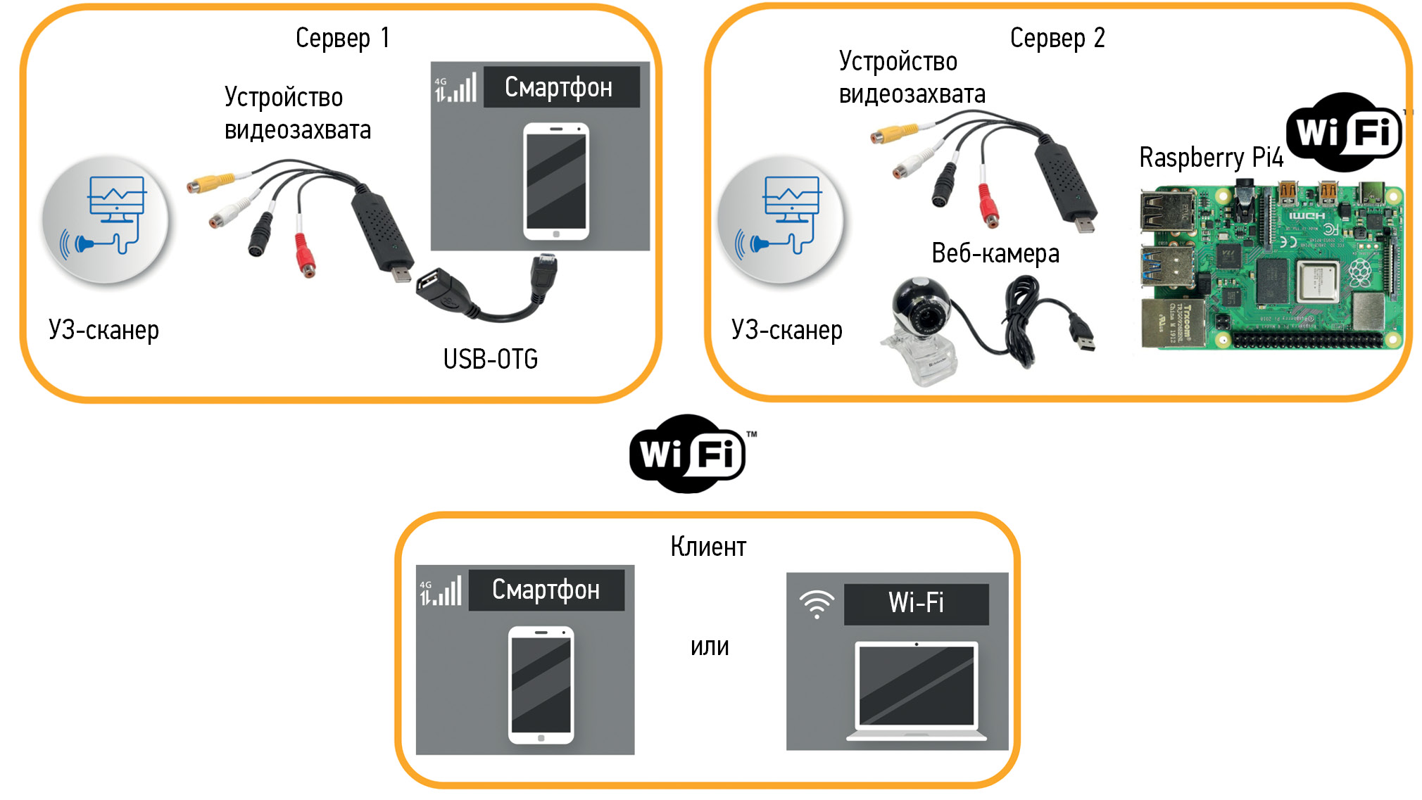
MATERIALS AND METHODS: This study focused on capturing ultrasound videos using external video recording devices connected via USB. Raspberry Pi single-board computers and Android smartphones have been used as platforms to host a tele-ultrasound server. Used software: VLC, Motion, and USB camera. A remote expert assessment was performed with mobile devices using the following software: VLC acted as a VLC server, Google Chrome for OS Windows 7 and OS Android was used in the remaining scenarios, and Chromium browser was installed on the Raspberry Pi computer.
OUTCOMES: The UTV007 chip-based video capture device produces better images than the AMT630A-based device. The optimum video resolution was 720×576 and 25 frames per second. VLC and OBS studios are considered the most suitable for a raspberry-based ultrasound system owing to low equipment and bandwidth requirements (0.64±0.17 Mbps for VLC; 0.5 Mbps for OBS studio). For Android phone OS, the ultrasound system was set with the USB camera software, although it required a faster network connection speed (5.2±0.3 Mbps).
CONCLUSION: The use of devices based on single-board computers and smartphones implements a low-cost tele-ultrasound system, which potentially improves the quality of studies performed through distance learning and consulting doctors. These solutions can be used in remote regions for “field” medicine tasks and other possible areas of m-health.
15-23
Systematic reviews
Low-dose computed tomography in COVID-19: systematic review
Аннотация
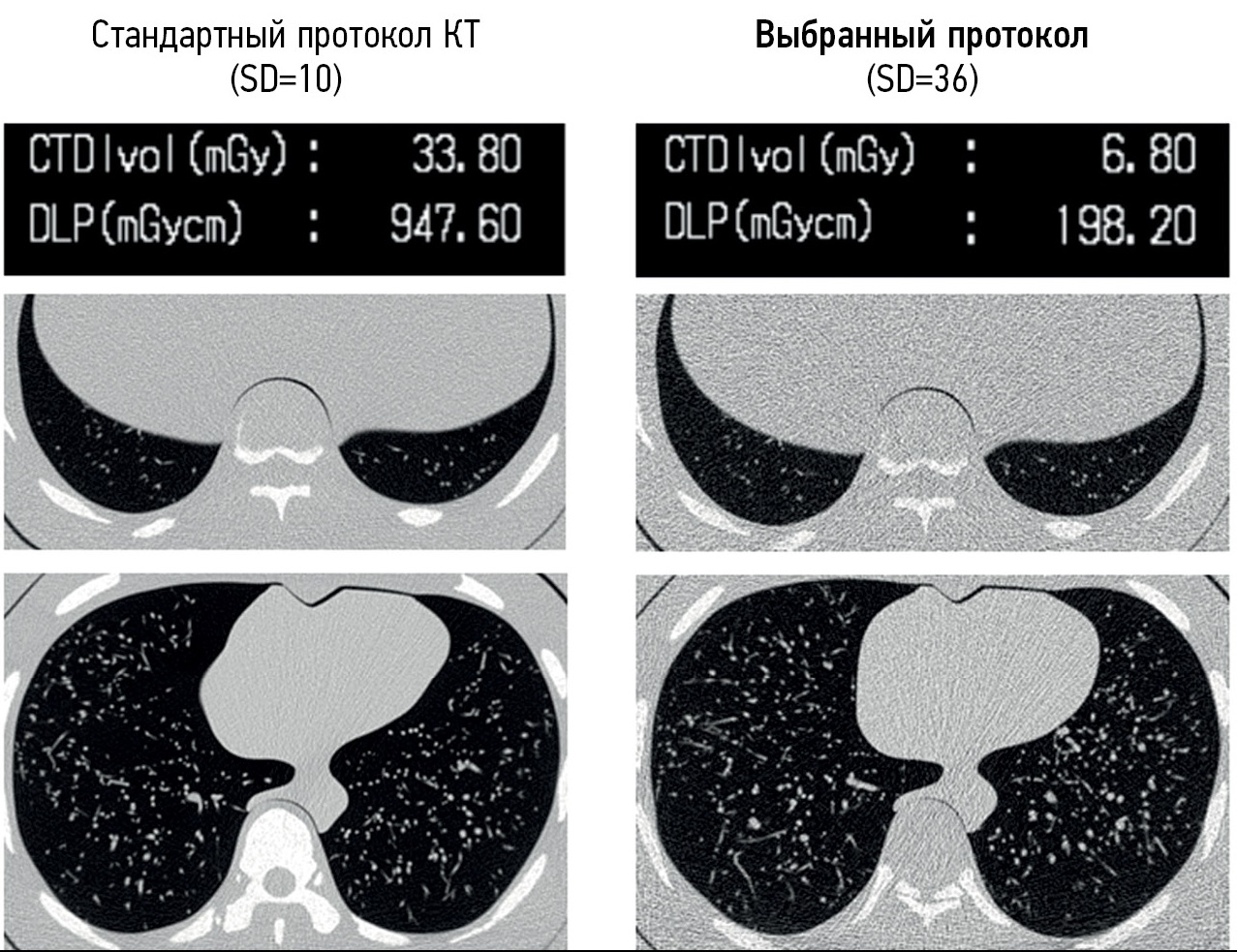
BACKGROUND: The increased number of computed tomography scans during the COVID-19 pandemic has emphasized the task of decreasing radiation exposure of patients, since it is known to be associated with an elevated risk of cancer development. The ALARA (as low as reasonably achievable) principle, proposed by the International Commission on Radiation Protection, should be adhered to in the operation of radiation diagnostics departments, even during the pandemic.
AIM: To systematize data on the appropriateness and effectiveness of low-dose computed tomography in the diagnosis of lung lesions in COVID-19.
MATERIALS AND METHODS: Relevant national and foreign literature in scientific libraries PubMed and eLIBRARY, using English and Russian queries “low-dose computed tomography” and “COVID-19,” published between 2020 and 2022 were analyzed. Publications were evaluated after assessing the relevance to the review topic by title and abstract analysis. The references were further analyzed to identify articles omitted during the search that may meet the inclusion criteria.
RESULTS: Published studies summarized the current data on the imaging of COVID-19 lung lesions and the use of computed tomography scans and identified possible options for reducing the effective dose.
CONCLUSION: We present techniques to reduce radiation exposure during chest computed tomography and preserve high-quality diagnostic images potentially sufficient for reliable detection of COVID-19 signs. Reducing radiation dose is a valid approach to obtain relevant diagnostic information, preserving opportunities for the introduction of advanced computational analysis technologies in clinical practice.
25-37
Reviews
Basic pulse sequences in the diagnosis of abdominal pathology
Аннотация
Magnetic resonance imaging is used for diagnosing abdominal and retroperitoneal space pathology, which allows visualizing focal or diffuse lesions in the parenchymal and hollow viscera with high diagnostic accuracy and reproducibility. Magnetic resonance imaging has advantages over computed tomography in the sensitivity and specificity of determining pathological changes in parenchymal organs, bile ducts and ducts of the pancreas, peritoneum, and retroperitoneal space.
The multiparametric protocol provides information about the mutual topography of organs and their structure and the functional state of tissues. This allows to move from structural to functional evaluation. In most cases, the standard abdominal protocol includes T1-weighted images, T2-weighted images, diffusion-weighted images, and magnetic resonance cholangiopancreatography. Depending on the objectives and patient’s condition, this protocol can be significantly reduced or supplemented.
Existing technical developments and achievements make it possible to simplify the scanning process and reduce the time for obtaining images while increasing the reproducibility of techniques in different healthcare institutions.
39-50
Case reports
Diseases and abnormalities of the nipple-areolar complex: a case report series
Аннотация
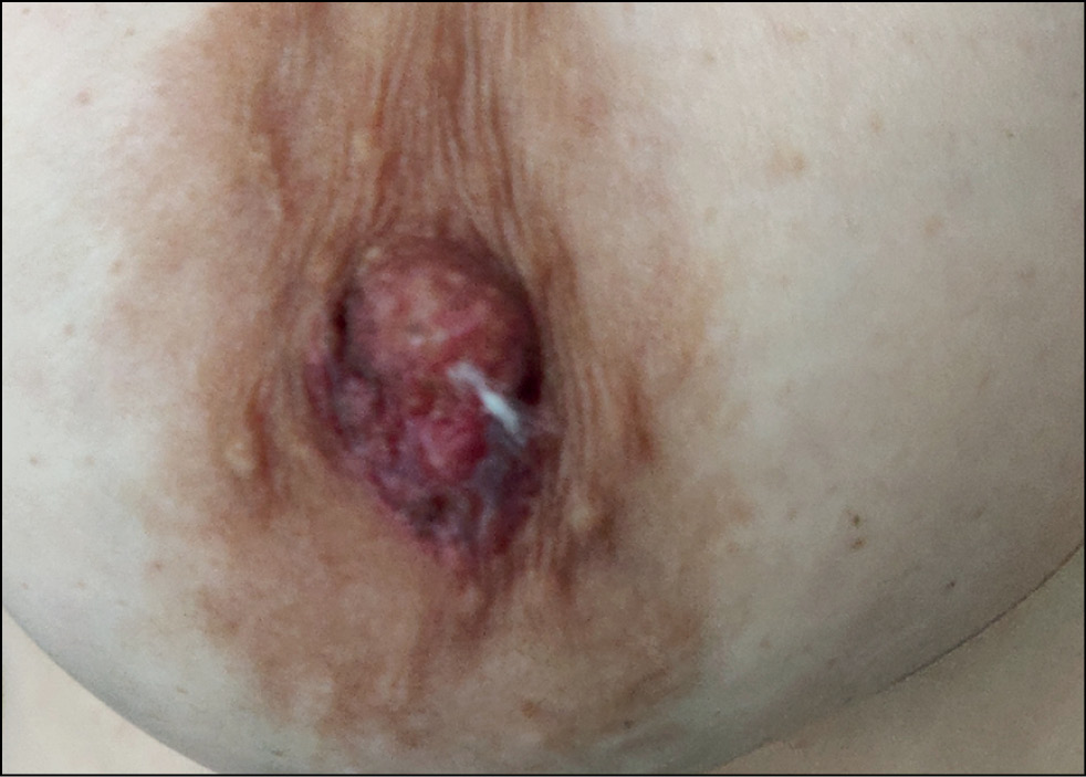
The nipple–areolar complex is a specific anatomical and histological structure. Normal structure and pathological process variabilities and the complexity of diagnostic imaging cause difficulties for radiologists and physicians. Breast magnetic resonance imaging is highly sensitive for structural features and nipple-areolar complex cancer detection. Magnetic resonance imaging is a useful diagnostic tool when mammography and ultrasound findings are inconclusive. It allows visualization of the retroareolar region, suitable for the diagnosis of papillomas, adenomas, Paget’s disease, ductal carcinoma in situ, and invasive ductal carcinoma.
This is a case report on identifying the pathology and anomalies of the nipple-areolar complex, which may benefit radiologists, gynecologists, and residents.
51-60
Magnetic resonance imaging in the diagnosis of necrosis of a pulled-through colon segment after abdomino-anal resection of the rectum for cancer

Аннотация
This study presents a case of necrosis of the pulled-through colon after abdomino-anal resection of the rectum, which was diagnosed by magnetic resonance imaging.
A 47-year-old man underwent laparoscopically assisted abdomino-anal resection of the rectum with reconstruction of a coloplasty pouch and transverse colostomy in the course of combination treatment for locally advanced rectal cancer. The postoperative period was complicated by the development of an inflammatory response syndrome. On postoperative day 3, contrast-enhanced magnetic resonance imaging revealed swelling of the 15-cm segment of pulled-through colon up to the coloanal anastomosis with sharply attenuated contrast enhancement, whereas rectoscopy showed no changes. On postoperative day 6, a magnetic resonance imaging scan revealed a defect in the anterior wall of the coloplasty pouch with a parietal aerocele, and rectoscopy showed signs of necrosis of the bowel wall. On postoperative day 10, the magnetic resonance imaging scan presented no changes. Because of increasing signs of inflammation, relaparotomy with anastomosis disconnection and resection of the necrotized bowel segment were performed.
Ischemia of the pulled-through colon after rectal surgery is a rare but serious complication. Our clinical case report demonstrates the potential of contrast-enhanced magnetic resonance imaging as a non-invasive method in case follow-up in patients with a complicated postoperative period for early diagnosis of ischemia and bowel wall defects, which helps to make the appropriate patient management plan.
61-69
Dynamic MRI in a COVID-19 patient: a case series
Аннотация
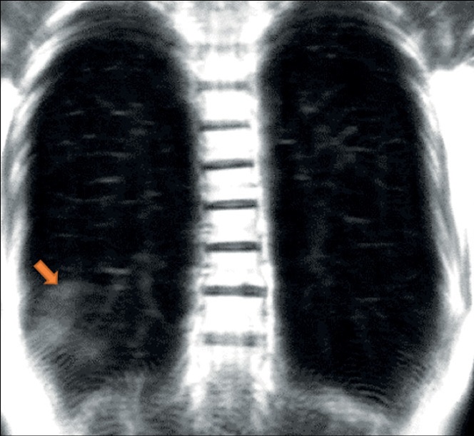
Extensive spread of the coronavirus disease (COVID-19) prompted an investigation of its diagnostic features. Acute viral pneumonia associated with COVID-19 has been described in detail using CT, radiography, and MRI. There is no data in the literature on the descriptive picture observed with dynamic MRI. Considering a comprehensive diagnostic approach, radiologists should know how to correctly recognize and interpret COVID-19 on MRI. This case series demonstrated the ability of dynamic MRI to detect the cloudy sky sign and distinguish it from consolidation in COVID-19 patients, thus presumably distinguishing between early or mild changes and a progressive clinical course. These changes in dynamic lung images on MRI can be recorded depending on the phase of the respiratory cycle. Thus, MRI, as a radiation-free tool that can be used to examine a patient with acute viral pneumonia COVID-19, can be useful in cases where access to computed tomography is limited and dynamic morphofunctional imaging is required.
71-79
Correspondence
Artificial intelligence in clinical physiology: How to improve learning agility
Аннотация
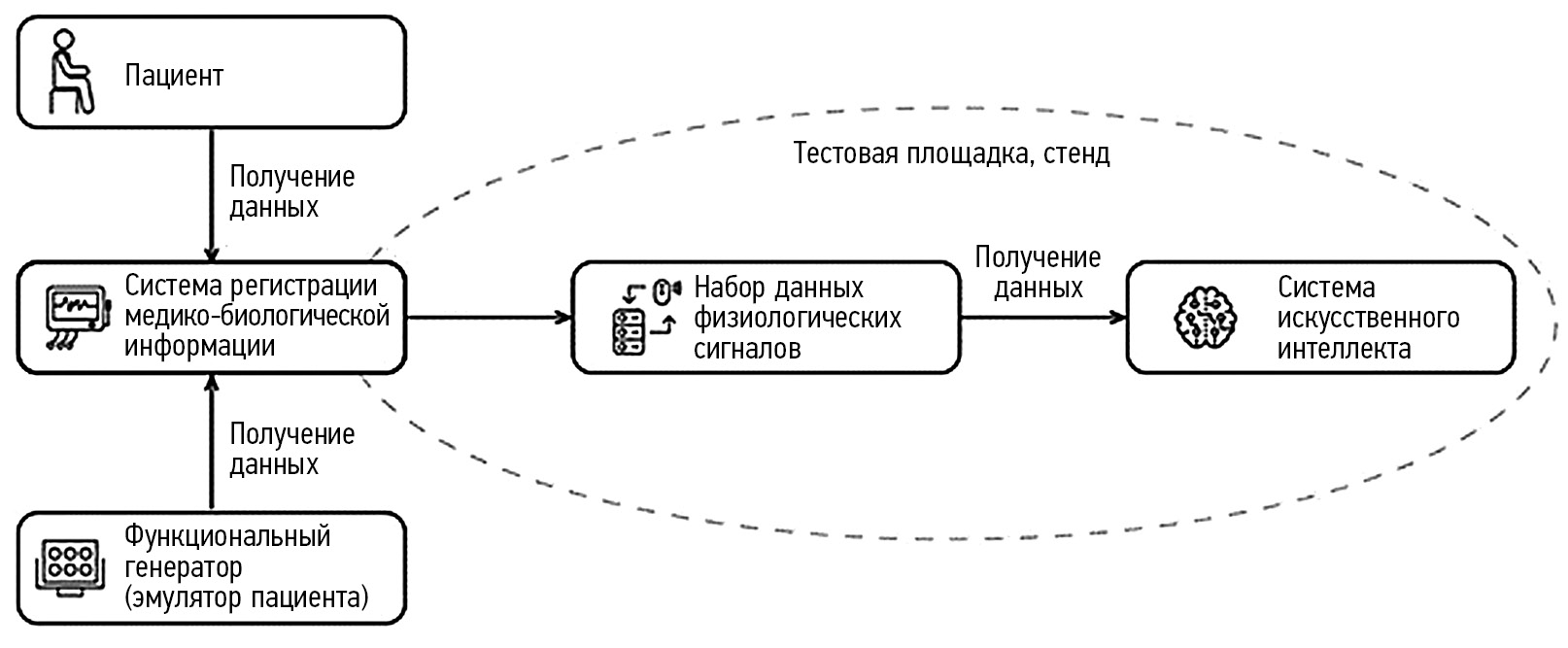
Clinical physiology involves a complete, comprehensive, multilateral study of the functions of both affected and healthy organs, which allows us to assess the compensatory capabilities of the body.
Artificial intelligence is increasingly being used in medicine, including in clinical physiology. This is facilitated by the increase in computing processing power, development of cloud services and datasets, and numerous scientific articles demonstrating the effectiveness and viability of such intelligent solutions.
Although the approach to medical dataset development is generally similar, there are a number of key features and significant differences in clinical physiology. Artificial intelligence systems in clinical physiology may be effectively trained and applied in practice by following the recommendations in this study.
The national standard of the Russian Federation GOST R 59921.9-2022, which has entered into force, is included in the set of standards “Artificial Intelligence systems in clinical medicine” and establishes additional requirements for data analysis algorithms and test methods of artificial intelligence systems used in the field of clinical physiology. A crucial feature of the created standard is its qualimetric type (i.e., it has a mandatory set of demonstration data).
Russia is one of the first countries to start developing quasi-metric standards worldwide, and 15 industry standards in the field of artificial intelligence (2 of them in medicine) will come into force this year.
81-88
Регистрационный номер и дата принятия решения о регистрации СМИ: серия ПИ № ФС 77 - 79539 от 09 ноября 2020 г.