

")


Том 2, № 3 (2021)
- Жылы: 2021
- ##issue.datePublished##: 15.10.2021
- Мақалалар: 12
- URL: https://jdigitaldiagnostics.com/DD/issue/view/3396
- DOI: https://doi.org/10.17816/DD.23

Original Study Articles
Electrocardiographic findings in COVID-19: analysis of tele-ECGs in Moscow ECG IT Center

Аннотация
BACKGROUND: Coronavirus disease (COVID-19) affects the cardiovascular system and the primary damage to the respiratory system involved in the pathological process. However, in the available literature, the electrocardiography (ECG) analyses are based only on small-sample studies and case reports, which determine the relevance of larger-scale studies to clarify the nature and prevalence of ECG abnormalities in subjects with confirmed coronavirus infection.
AIM: To determine the distribution of ECG changes in COVID-19 patients representing a non-selective population of Moscow residents.
MATERIALS AND METHODS: We performed a retrospective analysis of ECGs from 42,799 patients from March 10, 2020 to March 10, 2021 with a verified diagnosis of COVID-19 was performed. The study included patients admitted to Moscow clinical hospitals connected to the ECG IT Center. A standard 12-lead ECG was obtained and transmitted via an Internet connection to the server of the ECG IT Center, where the ECG interpretation was performed.
RESULTS: ECG changes were detected in 54% of patients. The most common cardiac arrhythmias were supraventricular extrasystole (12.6%) and atrial fibrillation (12.0%) reported in patients. Signs of the overloaded right heart were detected in 12.5% of cases, of which the ECG pattern of pulmonary embolism was confirmed in 485 patients (1.13%). Infarction ECG pattern was observed in 4.5% of patients, among which 3 cases of Brugada ECG pattern were reported. The incidence of ST-T changes was 2.2% of all study patients. Prolonged QT and QTc intervals were recorded in 540 patients (1.26%). In addition, individual cases of ventricular fibrillation, Frederick syndrome, and atrioventricular block of various degrees were reported.
CONCLUSION: The distribution of incidence of ECG changes in COVID-19 was shown based on the data obtained. The high incidence of atrial fibrillation, which is a risk factor for thromboembolic complications, was confirmed. Moreover, a significant prevalence of ECG patterns of overloaded right heart was shown, some are associated with pulmonary embolism. Other reported ECG changes were characterized by a significantly lower prevalence, which does not reduce their clinical significance. The data obtained may be used to improve COVID-19 patient management strategy in the future.
 235-248


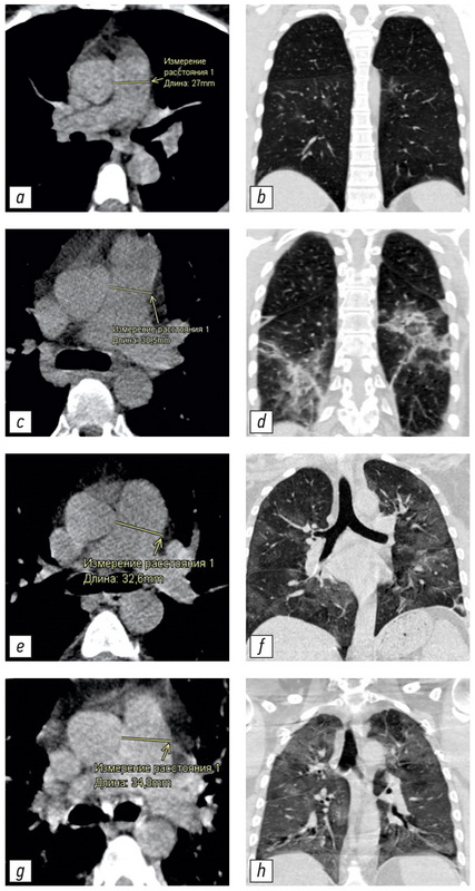
Changing of pulmonary artery diameter in accordance with severity of COVID-19 (assessment based on non-contrast computer tomography)
Аннотация
BACKGROUND: Computed tomography is the method of choice for assessing the volume of lung damage in viral pneumonia, including those associated with COVID-19. In addition, computed tomography can determine the main vessels size of the thorax. This allowed us to analyze the relationship between the severity of COVID-19 and the changes in the diameters of the pulmonary artery (PA) and ascending aorta (Ao). Dilation of the PA is a sign of pulmonary hypertension. The study of these patterns may be of clinical significance in determining the treatment tactics and prognosis of the course of COVID-19 disease.
AIM: To evaluate the relationship between PA diameter and the severity of the COVID-19 course in patients of different ages.
MATERIALS AND METHODS: This study is a single-centered, cross-section, continuous, uncontrolled study performed on a group of patients (n=511, 267 men, median 59 years, IQR 49.0–65.0, ages 31–84 years) who were treated in a temporary hospital to treat patient with COVID-19. During hospitalization all patients were examined by CT scan of the chest. All studies were carried out using a mobile CT scan system Airo TruCT (Stryker, USA). The degree of damage to the lung tissue was assessed using the CT volume scale 1–4. Measurement of the LA and Ao diameters was carried out using standard instruments of the radiologist’s CT workstation perpendicular to the long axis of the vessel.
RESULTS: The following statistically significant regularities were obtained: the detection of a dilated pulmonary artery (PA) and an increased PA/Ao ratio correlated to an increase in the degree of lung damage in COVID-19 (Kruskal-Wallis test, K-W p <0.001; median test, MT p <0.001), the diameter of the ascending aorta (Ao) significantly increases with the patient’s age (K-W p <0.001; MT p <0.001). An insignificant correlation between an increase in the diameter of the pulmonary artery (PA) and the patient’s age (K-W p=0.094; MT p=0.311) and an insignificant correlation between detection of a change in aortic (Ao) diameter and the degree of lung damage (K-W p=0.061; MT p=0.165) were shown. In groups with a severe course of the disease and a large volume of lung lesions (CT-3 and CT-4), a significantly greater number of patients with signs of pulmonary hypertension (detection of the dilated pulmonary artery: 29 mm and more) was shown for all age groups.
CONCLUSION: The study showed that PA dilatation and increased PA/Ao diameter ratio were significantly associated with increased pulmonary lesion volume in COVID-19 in all age groups.
249-260
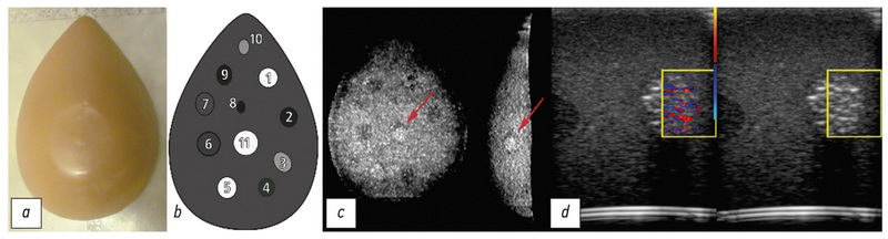
Doppler twinkling artifact observations: an open-access database of raw ultrasonic signals
Аннотация
BACKGROUND: Doppler twinkling artifact is a rapid change of colors seen in CFI-mode in the presence of kidney stones and calculi. Therefore, numerous researchers use the twinkling artifact as a diagnostic sign. However, this phenomenon is under-researched, because most assumptions concerning its causes are made based on pure visual observations of the scanner’s screen leaving the important steps of signal transformation hidden behind the “black box” curtains of ultrasound machines.
MATERIALS AND METHODS: Raw radiofrequency ultrasound signals were recorded in the phantom studies. The recorded echoes were received from objects that create the Doppler twinkling artifact and artificial blood vessels and soft tissues imitators. The data were collected between June 2016 and March 2021. Sonomed-500 with the 7.5 L38 and 3.4 C60 probes served as the research machine for the signal capture.
Data records: We present the database containing raw radiofrequency ultrasound signals from the beam former output of the research ultrasound machine. The dataset consists of CFI and B-mode echoes recorded from twinkling objects. Therefore, this database can be useful for those who test, develop and study ultrasound signal processing algorithms. Furthermore, the database is freely available online. The 10.5 GB database consists of echoes received from five phantoms. Raw radiofrequency signals were stored in the binary files; scanning parameters were stored in text files. The database is available at: https://mosmed.ai/datasets/ultrasound_doppler_twinkling_artifact.
Code availability: The public can visualize the database content with the specially written program TwinklingDatasetDisplay available at: https://github.com/Center-of-Diagnostics-and-Telemedicine/TwinklingDatasetDisplay.git.
Usage notes: The database can be used to test and develop signal-processing algorithms, such as wall filtration, velocity estimation, feature extraction, speckle reduction, etc. Furthermore, the public is free to share (copy, distribute, and transmit) and remix (adapt and do derivative works) the dataset considering appropriate credit is given.
261-276
Technical Reports
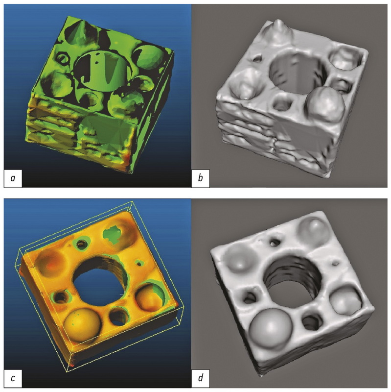
Evaluation of geometric deviations in rapid prototyped three-dimensional models created from computed tomography data
Аннотация
BACKGROUND: Computer-aided design and three-dimensional printing have been used in various clinical and fundamental medicine fields, especially in surgery. For example, in the preoperative period, the correspondence of printed products to the anatomy can play an important role in evaluating pathological changes and correction methods. However, determining dimensional deviations of printed models involves ethical and technical difficulties associated with defining a reference and taking many measurements, respectively. Therefore, we propose to use a geometric object with known dimensions as a reference and estimate linear deviations using the Iterative Closest Point algorithm for each of the vertices of the prototyped polygonal mesh.
AIMS: To evaluate the geometric deviations associated with creation of bone-like physical objects from computed tomography data using computer-aided design and additive manufacturing.
MATERIALS AND METHODS: The source object was created using the FreeCAD application; Blender and Meshmixer software was used for polygon meshes correction and transformation. The 3D printing was carried out on an Ender-3 printer with copper-impregnated polylactide plastic BFCopper. Scanning was performed using a 128-slice tomograph Philips Ingenuity CT. A series of tomographic images were processed in 3DSlicer software to create virtual models by semiautomatic segmentation with threshold values of 500 HU, 0 HU, −500 HU, −750 HU, and manual segmentation. Reproduced and reference polygon meshes were compared using the Iterative Closest Point algorithm in CloudCompare software.
RESULTS: The volume of reproduced models exceeded the volume of respective reference models by 1%–27%. The average point cloud linear deviation values of reproduced models from the reference ones were 0.03–0.41 mm. A significant correlation between integral sums of linear deviations and changes in the volume of reproduced models was shown using Spearman's rank correlation coefficient (ρ = 0.83; temp = 5.27, p=0.05).
CONCLUSION: The geometry of the reproduced object changes inevitably, while the linear deviations depend more on the chosen segmentation method than on the overall size of the model or its structures. The manual segmentation method can lead to greater linear deviations, though it saves all the necessary anatomical structures.
277-288
Systematic reviews
Objective criteria for MRI evaluation of the effectiveness of treatment of bone metastases in patients with prostate cancer and breast cancer: systematic review and meta-analysis
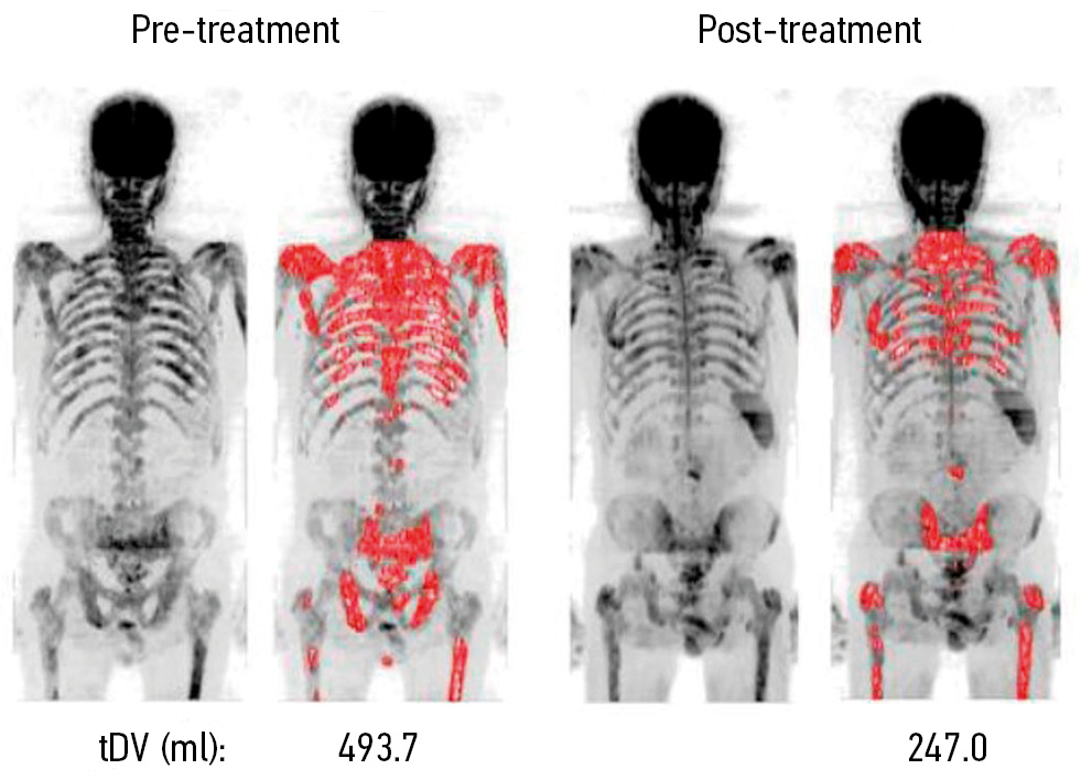
Аннотация
BACKGROUND: The possibility of a personalized approach to the treatment of metastatic prostate cancer and breast cancer requires objective methods for the evaluation of the response of foci treatment in the skeleton. The proven high efficiency of MRI in detecting bone metastases, in combination with the absence of ionizing radiation, has laid the groundwork for using this method in monitoring the treatment course based on objective criteria for evaluation of the therapeutic outcome.
AIM: To assess the possibilities of quantitative and semi-quantitative parameters of MRI-evaluation of treatment efficacy (radiation, chemotherapy, hormone therapy, and targeted therapy) of bone metastases that were used in prostate and breast cancer clinical trials.
MATERIALS AND METHODS: We searched the databases Embase, PubMed, Cochrane Central Register of Controlled Trials (CENTRAL), eLibrary until April 1, 2021, using the following keywords: magnetic resonance imaging, MRI, DWI, treatment response, prostate or breast cancer, and bone metastasis. We only included studies related to the MRI-evaluation of treatment efficacy of any type of therapeutic intervention (with the exception of surgery) for metastatic skeletal lesions in this review.
RESULTS: We selected and analyzed 11 out of 312 sources found as a result of the search. It allowed us to identify four groups of objective MRI criteria for evaluating the therapeutic effect in metastatic bone lesions in patients with prostate and breast cancer, including the dynamics of sizes, signal intensity on DWI, ADC, and tumor total diffusion volume (tDV). Changes in these quantitative and semi-quantitative indicators, with only one exception, had the same direction, although they differed in numerical values. A random-effects model was used for analysis considering the presence of statistically significant heterogeneity (p <0,1 for χ2 test; I2 >40%),. The change in ADC as a result of treatment averaged +0.35 [+0.12; +0.49] ×10−3 mm2/s, with average values of ADC before treatment ― 0.83 [0.71; 1.03] ×10−3 mm2/s, after treatment ― 1.18 [0.83; 1.49] ×10−3 mm2/s.
CONCLUSION: MRI is an informative technique for the objective evaluation of the response of bone metastases to therapy in patients with prostate cancer and breast cancer based on quantitative and semi-quantitative parameters. It has significant potential as a diagnostic test instrument for monitoring the effectiveness of treatment of metastatic skeletal lesions.
289-300
Role of chest MRI for the diagnosis of malignant pulmonary nodules: a systematic review and a meta-analysis
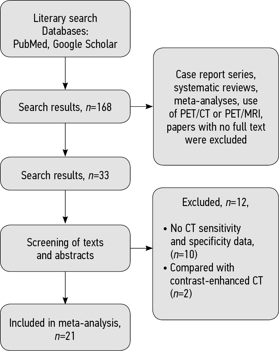
Аннотация
AIM: To evaluate the ability of magnetic resonance imaging (MRI) of the chest to detect malignant pulmonary nodules compared to compute tomography (CT).
MATERIALS AND METHODS: We searched the following databases with the final date of search on April 7th, 2021: PubMed, Google Scholar. We selected studies according to the inclusion and exclusion criteria that assessed the detection of malignant lung nodules by MRI and CT and included information about sensitivity and specificity. Method of the analysis and data grouping was chosen with regard to statistical heterogeneity of the studies included in the analysis. We used the χ2 test and I2 statistic to evaluate the heterogeneity.
RESULTS: We selected 168 articles for the systematic review from the PubMed and Google Scholar databases. We included 21 studies on 1,188 patients in the meta-analysis and revealed statistically significant heterogeneity (р<0,00001 for χ2 test; I2=99%) for sensitivity and specificity. Hence, we used a random-effect model for further analysis. As a result, values of sensitivity for detection of pulmonary nodules with MRI of 70.4%–100%, specificity ― from 60.6% to 100%.
CONCLUSIONS: Thus, MRI has sufficient sensitivity and specificity for detecting malignant pulmonary nodules primarily discovered with CT.
301-312
Reviews
STARD 2015 guidelines for reporting diagnostic accuracy studies: explanation and elaboration. Translation to Russian
Аннотация
Diagnostic accuracy studies are, like other clinical studies, at risk of bias due to shortcomings in design and conduct, and the results of a diagnostic accuracy study may not apply to other patient groups and settings. Readers of study reports need to be informed about study design and conduct, in sufficient detail to judge the trustworthiness and applicability of the study findings. The STARD statement (Standards for Reporting of Diagnostic Accuracy Studies) was developed to improve the completeness and transparency of reports of diagnostic accuracy studies. STARD contains a list of essential items that can be used as a checklist, by authors, reviewers and other readers, to ensure that a report of a diagnostic accuracy study contains the necessary information. STARD was recently updated. All updated STARD materials, including the checklist, are available at http://www.equator-network.org/reporting-guidelines/stard. Here, we present the STARD 2015 explanation and elaboration document. Through commented examples of appropriate reporting, we clarify the rationale for each of the 30 items on the STARD 2015 checklist, and describe what is expected from authors in developing sufficiently informative study reports.
This article is the reprint with Russian translation of the original that can be observed here: Cohen JF, Korevaar DA, Altman DG, et al. STARD 2015 guidelines for reporting diagnostic accuracy studies: explanation and elaboration. BMJ Open 2016;6:e012799. doi: 10.1136/bmjopen-2016-012799
313-342
Dual-energy computed tomography for head and neck cancer
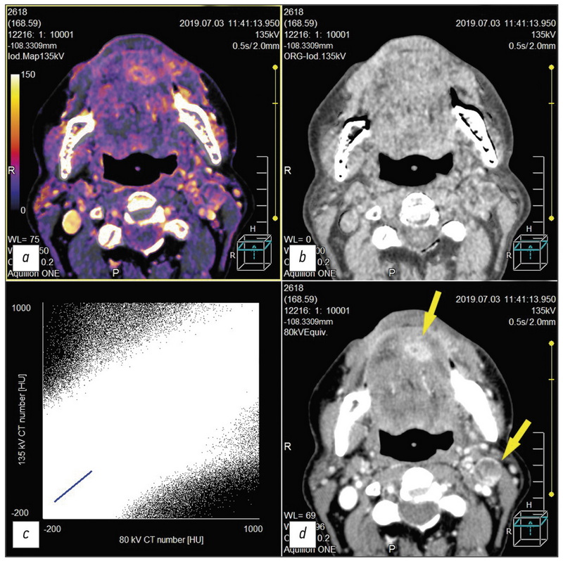
Аннотация
This study reviewed the head and neck cancer diagnosis publications using dual-energy computed tomography (DECT). The qualitative and quantitative analysis of the data was DECT obtained using intravenous contrast enhancement for localized tumors, which shows the importance of constructing iodine maps for obtaining additional diagnostic information. Including the article is described aspects of improving visualization of the oropharyngeal region against the background of artifacts from dental implants. Several research articles highlight the current state of the issue and the role of post-processing of “raw data” DECT, obtaining a range of monochromatic images of a tumor and other pathological changes in the head and neck region in the article. Several learned treatises were also reflected. DECT with intravenous contrast enhancement and routine computed tomography to reduce radiation exposure to patients were compared particularly due to the possibility of obtaining virtual native diagnostic images from a contrasting series of DECT volumes during post-processing. In addition, this review also includes references to works that highlight the development of DECT as the method. Finally, the physical principles underlying DECT and the prospects for the development of the method are briefly represented.
343-355
On the issue of ethical aspects of the artificial intelligence systems implementation in healthcare
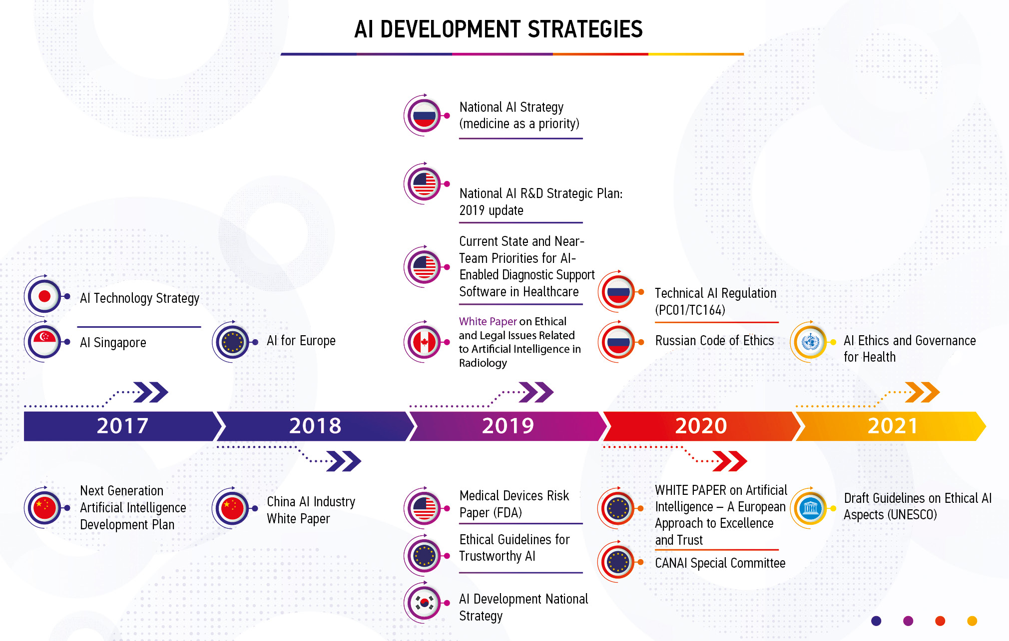
Аннотация
The article analyzes ethical issues inherent to different life-cycle stages of artificial intelligence systems and provides up-to-date information about global and domestic trends in this area. The international and national experience concerning ethical issues of artificial intelligence systems use in healthcare is described. In addition, the international and national strategies for the development of artificial intelligence in healthcare are analyzed focusing on the national development; and the main trends, similarities, and differences between strategies are identified. Furthermore, ethical components of the process of clinical trials to evaluate the safety and efficacy of artificial intelligence systems in Russia are described. Domestic state-of-the-art and globally unique experience in technical regulation of artificial intelligence systems are shown on unification papers and standardization of requirements for the development, testing, and operation of artificial intelligence systems in healthcare are presented, and unparalleled Russian experience in terms of certification requirements for artificial intelligence-based medical devices is demonstrated.
The article also summarizes the main conclusions and emphasizes the importance of a strong successful healthcare system based on artificial intelligence technologies that build trust and compliance with ethical standards.
356-368
Methods of medical visualization and thermal ablation as a new approach to treatment of hyperparathyroidism

Аннотация
The pathologies of parathyroid glands are widespread among endocrine system diseases, excluding diabetes and thyroid pathology. There are only two methods that are used to treat hyperparathyroidisms, such as surgery and conservative therapy. However, transracial thermal destruction methods (ablation) have recently appeared in clinical practice. The methods have good precision and connect with physical phenomena, such as interaction laser, radiofrequency, microwave, and HIFU irradiation with bio substance. The review is dedicated to critically analyze the modern methods for local thermal destruction of the hyper-functioning parathyroid glands. The review includes data from randomized clinical trials from 2012 to 2021. The studies were from Google Scholar and Pubmed with a total number of 1,938 patients (laser ablation ― 216 patients, radiofrequency ablation ― 225, microwave ablation ― 1467, high-density ultrasound ablation ― 30 patients). Recommendations methods of thermal destruction application were obtained during the review. Furthermore, we have designed some algorithms for hyperparathyroidism treatment. Moreover, thermal destruction methods were observed. There are four modern methods of thermal destruction which have been analyzed like alternatives to surgery. Each of them has advantages and disadvantages, its profile of safety and effectiveness. After processing information from a proven database, the most popular among specialists is methods of microwave ablation. However, laser ablation is more effective than other ways.
369-385
Emerging techniques in atherosclerosis imaging
Аннотация
Atherosclerosis is a chronic immunomodulated disease that affects multiple vascular beds and results in a significant worldwide disease burden. Conventional imaging modalities focus on the morphological features of atherosclerotic disease such as the degree of stenosis caused by a lesion. Modern CT, MR and positron emission tomography scanners have seen significant improvements in the rapidity of image acquisition and spatial resolution. This has increased the scope for the clinical application of these modalities. Multimodality imaging can improve cardiovascular risk prediction by informing on the constituency and metabolic processes within the vessel wall. Specific disease processes can be targeted using novel biological tracers and “smart” contrast agents. These approaches have the potential to inform clinicians of the metabolic state of atherosclerotic plaque.
This review will provide an overview of current imaging techniques for the imaging of atherosclerosis and how various modalities can provide information that enhances the depiction of basic morphology.
This publication is the reprint with Russian translation from original: Syed MB, Fletcher AJ, Forsythe RO, Kaczynski J, Newby DE, Dweck MR, van Beek EJ. Emerging techniques in atherosclerosis imaging. Br J Radiol. 2019;92(1103):20180309. doi: 10.1259/bjr.20180309.
386-409
Editorials
Radiotheranostics is here to help

Аннотация
The COVID-19 pandemic did not diminish interest in radiotheranostics. However, the demand for visualization of pathological processes using cross-sectional and hybrid imaging (CT, MRI, SPECT, and PET) is increased. Over the past 15 months, the world has seen an exponential increase in investment in new radiopharmaceuticals for radiotheranostics. The list of antibodies and ligands labeled with "medical" radioactive isotopes is expanding as the molecular mechanisms of regulation and implementation of metabolic processes become clearer. The range of diagnostic and therapeutic radioactive isotopes is also expanding, ultimately increasing the range and availability of radiotherapy in nuclear medicine centers worldwide. It is necessary to unite the efforts of physicists, radiopharmacists, chemists, biologists, doctors, and mathematicians to develop radio technology. Usage and improvement of personalized dosimetry for planning radionuclide therapy is also a priority. For example, the International Foundation Oncidium helps with information and exchange of experience, while the international diagnostic study NOBLE increases the availability and reduces the cost of PSMA receptor scintigraphy. An association for the development of theranostics was created to intensify the integration renewal of nuclear medicine.
410-416
Регистрационный номер и дата принятия решения о регистрации СМИ: серия ПИ № ФС 77 - 79539 от 09 ноября 2020 г.