

")


Vol 4, No 2 (2023)
- Year: 2023
- Published: 12.07.2023
- Articles: 11
- URL: https://jdigitaldiagnostics.com/DD/issue/view/4600
- DOI: https://doi.org/10.17816/DD.42

Original Study Articles
Double-reading mammograms using artificial intelligence technologies: A new model of mass preventive examination organization
Abstract
BACKGROUND: In recent years, the availability of medical datasets and technologies for software development based on artificial intelligence technology has resulted in a growth in the number of solutions for medical diagnostics, particularly mammography. Registered as a medical device, this program can interpret digital mammography, significantly saving time, material, and human resources in healthcare while ensuring the quality of mammary gland preventive studies.
AIM: This study aims to justify the possibility and effectiveness of artificial intelligence-based software for the first interpretation of digital mammograms while maintaining the practice of a radiologist’s second description of X-ray images.
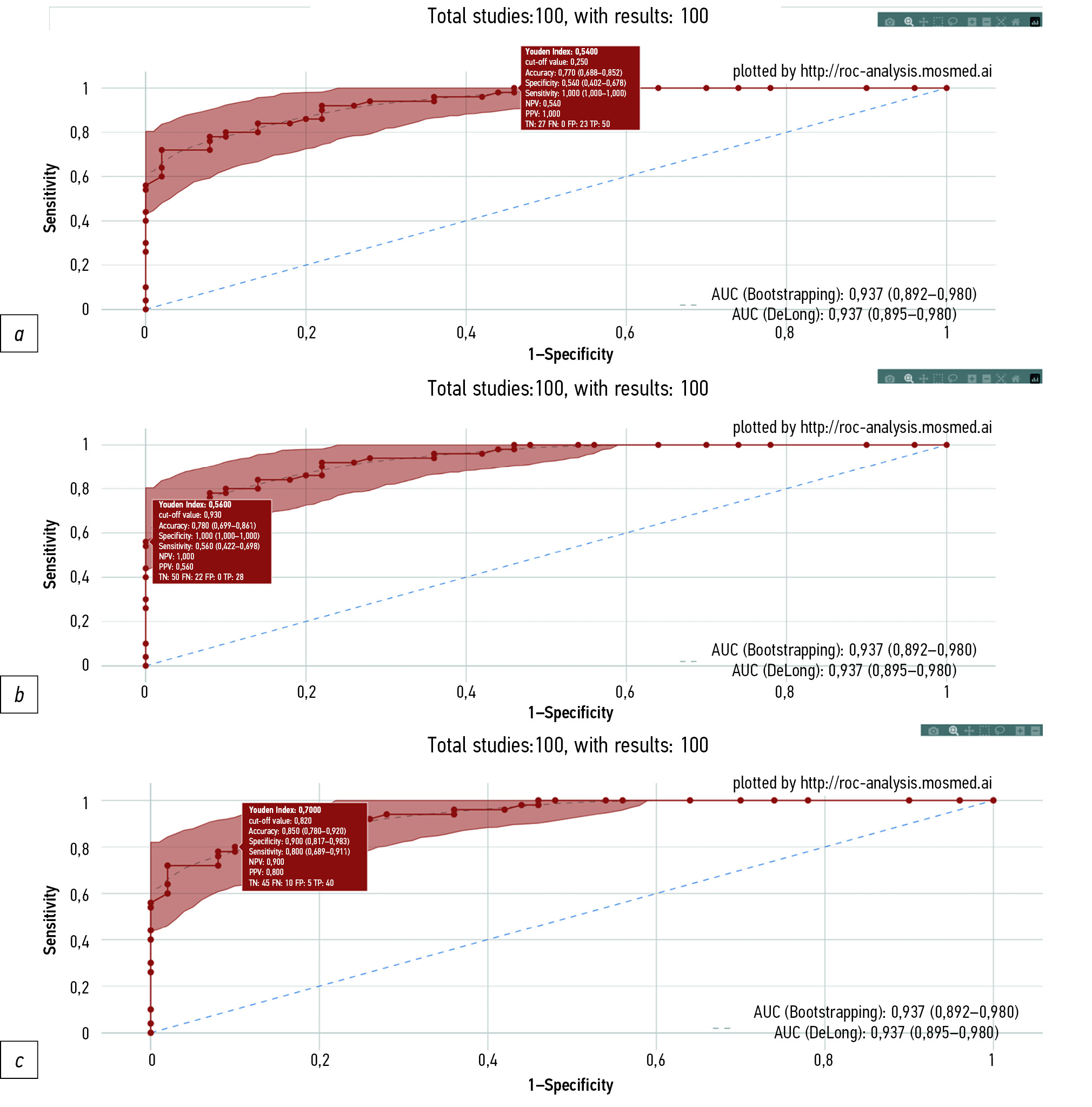
MATERIALS AND METHODS: A dataset of 100 digital mammography studies (50 — “absence of target pathology” and 50 ― “presence of target pathology,” with signs of malignant neoplasms) was processed by software based on artificial intelligence technology that was registered as a medical device in the Russian Federation. Receiver operating characteristic analysis was performed. Limitations of the study include the values of diagnostic accuracy metrics obtained for software based on artificial intelligence technology versions, relevant at the end of 2022.
RESULTS: When set to 80.0% sensitivity, artificial intelligence specificity was 90.0% (95% CI, 81.7–98.3), and accuracy was 85.0% (95% CI, 78.0–92.0). When set to 100% specificity, artificial intelligence demonstrated 56.0% sensitivity (95% CI, 42.2–69.8) and 78.0% accuracy (95% CI, 69.9–86.1). When the sensitivity was set to 100%, the artificial intelligence specificity was 54.0% (95% CI, 40.2–67.8), and the accuracy was 77.0% (95% CI, 68.8–85.2). Two approaches have been proposed, providing an autonomous first interpretation of digital mammography using artificial intelligence. The first approach is to evaluate the X-ray image using artificial intelligence with a higher sensitivity than that of the double-reading mammogram by radiologists, with a comparable level of specificity. The second approach implies that artificial intelligence-based software will determine the mammogram category (“absence of target pathology” or “presence of target pathology”), indicating the degree of “confidence” in the obtained result, depending on the corridor into which the predicted value falls.
CONCLUSIONS: Both proposed approaches for using artificial intelligence-based software for the autonomous first interpretation of digital mammograms can provide diagnostic quality comparable to, if not superior to, double-image reading by radiologists. The economic benefit from the practical implementation of this approach nationwide can range from 0.6 to 5.5 billion rubles annually.
 93-104


Analysis of the diagnostic and economic impact of the combined artificial intelligence algorithm for analysis of 10 pathological findings on chest computed tomography
Abstract
BACKGROUND: Artificial intelligence technology can help solve the significant problem of missed findings in radiology studies. An important issue is assessing the economic benefits of implementing artificial intelligence.
AIM: To evaluate the frequency of missed pathologies detection and the economic potential of artificial intelligence technology for chest computed tomography compared and validated by experienced radiologists.
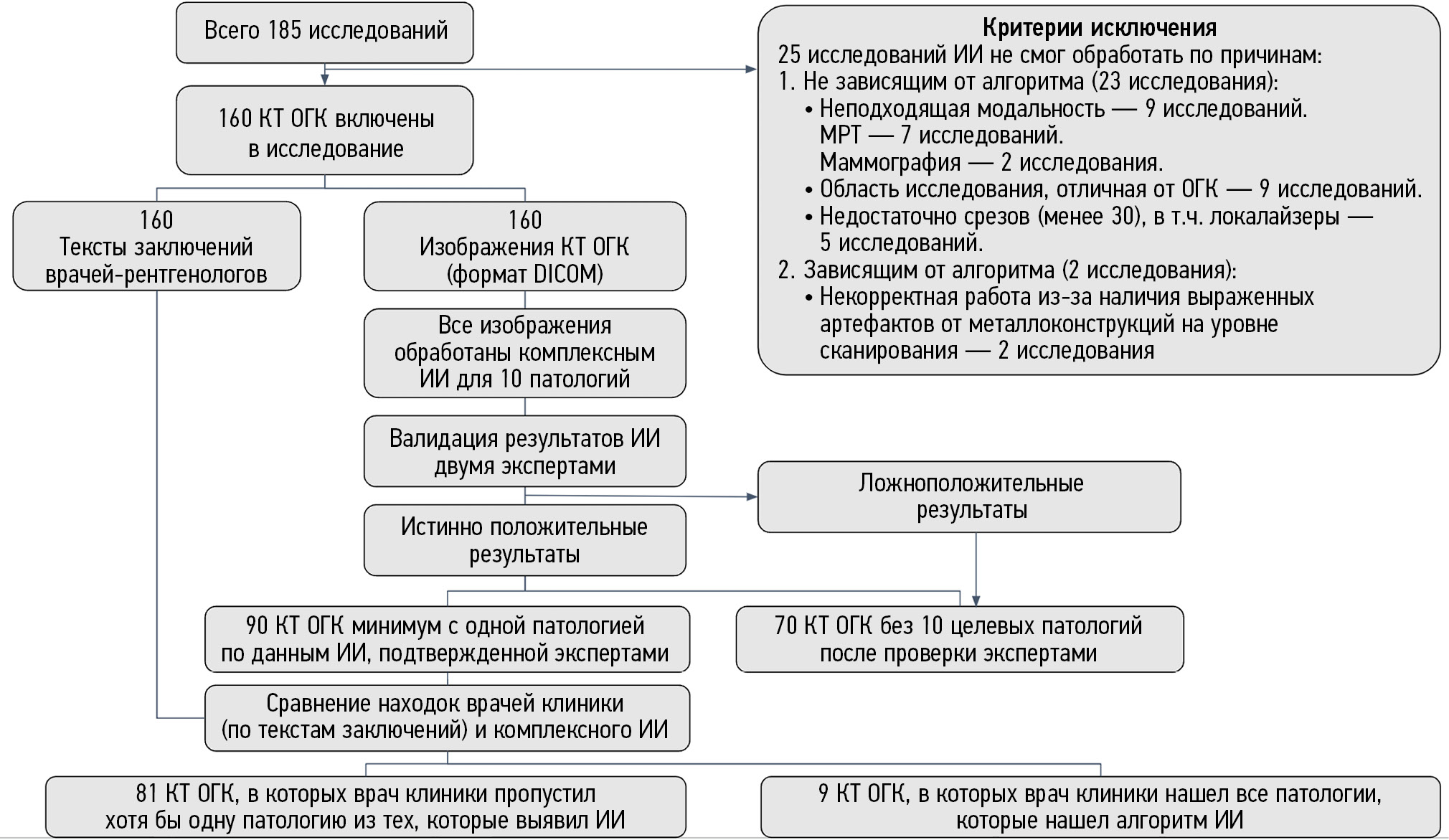
MATERIALS AND METHODS: This was an observational, single-center retrospective study. The study included chest computed tomography without IV contrast from June 1 to July 31, 2022, in Clinical Hospital in Yauza, Moscow. The computed tomography was processed using a complex artificial intelligence algorithm for 10 pathologies: pulmonary infiltrates, typical for viral pneumonia (COVID-19 in pandemic conditions); lung nodules; pleural effusion; pulmonary emphysema; thoracic aortic dilatation; pulmonary trunk dilatation; coronary artery calcification; adrenal hyperplasia; and osteoporosis (vertebral body height and density changes). Two experts analyzed computed tomography and compared results with artificial intelligence. Further routing was determined according to clinical guidelines for all findings initially detected and missed by radiologists. The hospital price list determined the potential revenue loss for each patient.
RESULTS: From the final 160 computed tomographies, the artificial intelligence identified 90 studies (56%) with pathologies, of which 81 (51%) were missing at least one pathology in the report. The “second-stage” lost potential revenue for all pathologies from 81 patients was RUB 2,847,760 ($37,251 or CNY 256,218). Lost potential revenue only for those pathologies missed by radiologists but detected by artificial intelligence was RUB 2,065,360 ($27,017 or CNY 185,824).
CONCLUSION: Using artificial intelligence as an “assistant” to the radiologist for chest computed tomography can dramatically minimize the number of missed abnormalities. Compared with the normal model without artificial intelligence, using artificial intelligence can provide 3.6 times more benefits. Using advanced artificial intelligence for chest computed tomography can save money.
105-132
Datasets
Creation of a training and test dataset with the disposition and transposition of overlaying electrocardiographic electrodes when recording electrocardiograms-12
Abstract
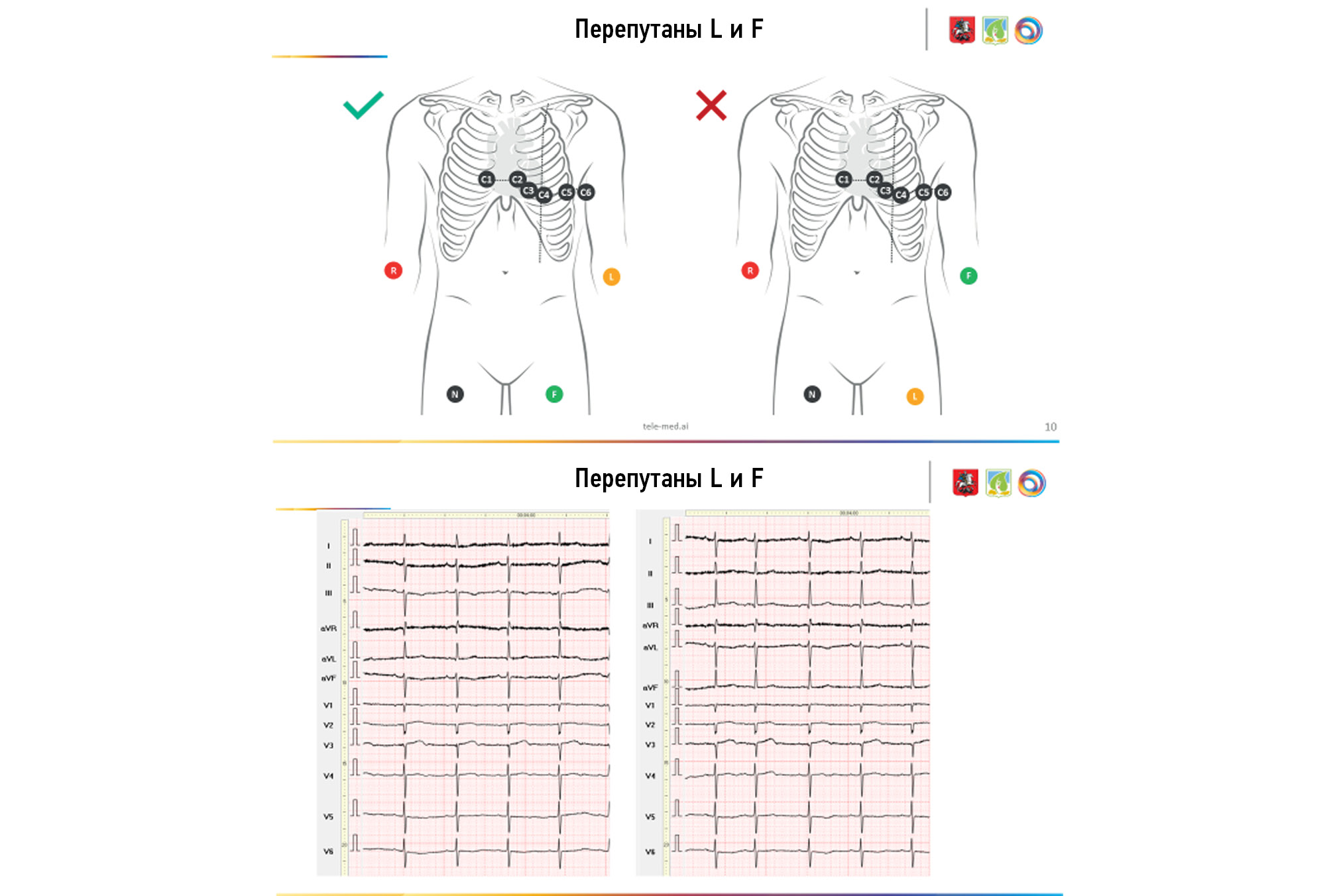
BACKGROUND: Electrocardiography is one of the simplest, most widely used, inexpensive, and informative methods in functional diagnostics; yet, if performed poorly, its diagnostic value is sharply reduced. Several attempts were made to systematize errors and deviations in electrode application, but all concerned the most common options (rearrangement of red and yellow electrodes, yellow and green electrodes, and chest electrodes — above or below the standard scheme).
AIM: To create an electrocardiogram dataset with different options for transpositions and dispositions of electrodes during electrocardiogram recording.
MATERIALS AND METHODS: The study included patients aged 18–75 years (27 males and 22 females). All patients provided informed consent for electrocardiogram registration. During one visit, the cardiogram was recorded on the device “Modular system for recording and remote transmission of electrocardiograms (EASY ECG)” for each patient.
RESULTS: In all, 488 electrocardiograms were recorded in 49 patients. The results obtained indicate a significant variability of the electrocardiogram pattern. Visual analysis of the electrocardiograms revealed no difficulties in determining the transposition associated with rearranging the leads on the arms (RY) in the thoracic C1–C2. The placement of thoracic electrodes in contact cheek-to-cheek dispositions with the transfer of thoracic leads above or below two intercostals was reliably determined compared with the Wilson scheme. The transpositions of the yellow and green limb electrodes and the change in the position of the thoracic ones when they are “lined up” in a straight line, “bullied” between the ribs (curved), and confused in places C5 and C6 are difficult to determine even when comparing two cardiograms next to each other, with the correct and transpositional superposition of the electrodes. The initial changes on the electrocardiograms, physique type, breast size, or the presence of an implant most likely determine it.
CONCLUSION: An electrocardiography dataset was obtained using various electrode dislocation variants. The dataset consists of a series of electrocardiograms obtained for each patient with several electrode placement options and contains both normal and pathological electrocardiograms.
133-141
Technical Reports
Problems of personnel irradiation in modern medical technologies
Abstract
BACKGROUND: The widespread use of radiation sources in medical practice (cardio-endovascular surgery, endoscopy, traumatology, urology, neurosurgery, dentistry, and radioisotope diagnostics departments) leads to irradiation of the lens of the eye and the skin of the hands. The introduction of new recommendations by the IAEA to reduce the limit of the annual equivalent dose to the lens (20 mSv) has led to an inaccurate dose assessment based on the effective dose.
AIM: To analyze approaches and assess equivalent doses of irradiation of the lens of the eye and skin of the hands of medical personnel during various diagnostic studies under the influence of X-rays and radiopharmaceuticals studies and to compare the results obtained with previously published data.
MATERIALS AND METHODS: Thermo-luminescent dosimetry was used. Dose assessment was performed by cardio-endovascular surgery, endoscopy, isotope diagnostics, dentistry, and urology personnel.
RESULTS: The estimated annual equivalent doses to the lens of the eye for doctors of cardio-endovascular surgery departments, in most cases, ranging 35–90 mSv, 6–19 mSv for the average medical staff (in some cases, the doctor [≤225 mSv] and the nurse [≤180 mSv]) and 4.5–9 mSv for the staff of the department of radioisotope diagnostics. The annual calculated equivalent doses to the skin of the hands for cardio-endovascular surgery personnel were 17–100 and 24–220 mSv for the staff working with radiopharmaceuticals. It is shown that the use of an estimate of the average dose per operation by cardio-endovascular surgery doctors, as a rule, inevitably leads to an excess of the equivalent dose to the lens of the eye after a certain number of operations.
CONCLUSION: When a certain number of operations are exceeded (100–200), equivalent doses to the eye’s lens in cardio-endovascular surgery doctors above 20 mSv per year can be formed. At current radiation levels, a lesion of the eye’s lens was found in a cardio-endovascular surgery doctor. The results indicate the need for further dosimetric measurements and epidemiological studies, based on which recommendations for radiation protection of the eye’s lens and the skin of the hands of medical personnel working in low-intensity, scattered, gamma X-ray radiation can be developed.
142-155
Systematic reviews
COVID-19-related cardiac lesion: The questions of pathogenesis and diagnostics
Abstract
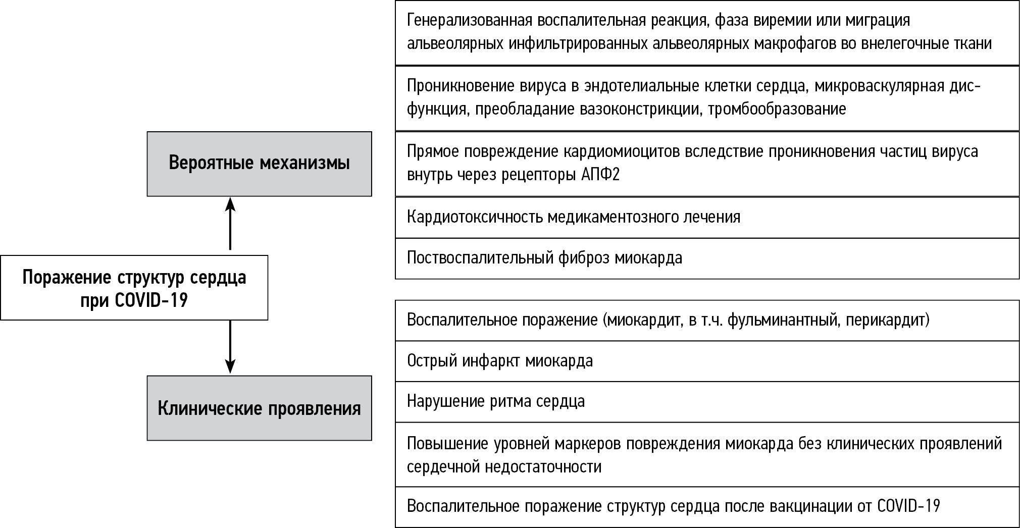
Coronavirus infection is still a topic of interest in the medical community today. Among the heterogeneous clinical manifestations of this disease, lesions of cardiac structures often occur. They are mainly inflammatory in nature and can be acute or delayed. Aside from myocarditis, coronavirus infection can induce cardiac injuries, including acute coronary syndrome, thromboembolic events, heart failure, and heart rhythm disturbances. It is well known that the prognosis for patients with cardiac lesions significantly worsens; timely diagnosis and treatment initiation play an important role in preventing severe complications.
This review presents the most recent literature data on the pathogenesis of cardiac lesions in COVID-19 patients and discusses the rational diagnosis of this pathology using modern techniques, such as laboratory, functional imaging (cardiac magnetic resonance is the most important of these), and invasive ones. It is now established that diagnosing myocarditis caused by coronavirus infection differs fundamentally from diagnosing other types of myocarditis. Furthermore, the main aspects of inflammatory heart lesions associated with COVID-19 vaccination are discussed, as this complication occurs more frequently than is commonly believed. It is often used as a rationale for refusing vaccination; however, this decision may severely affect the individual and the population.
156-169
Evaluation of fetal absorbed doses from computed tomography examinations of pregnant patients: A systematic review
Abstract
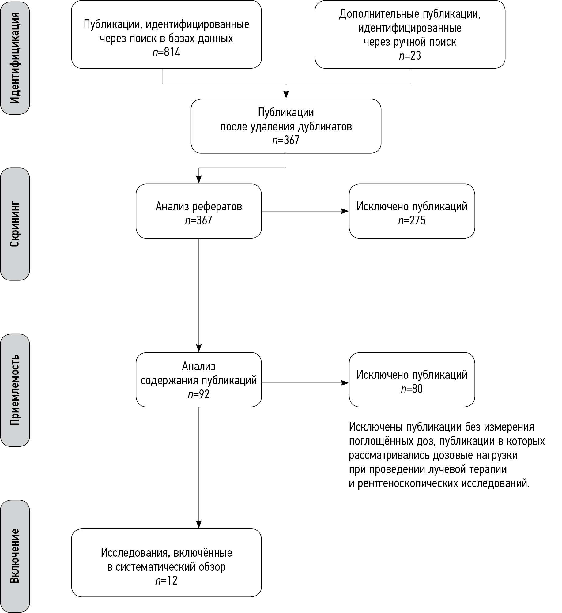
BACKGROUND: Currently, no systematic data are available on fetal radiation exposure as a result of radiographic studies during pregnancy. Consequently, there are no approved methods of its calculation that can be used in clinical practice. It is especially relevant for computed tomography scans as it is a widely used and highly informative method of diagnostic imaging associated with high exposure levels.
AIM: to systematize currently available data on radiation dose absorbed by the fetus from computed tomography scans in pregnant women.
MATERIALS AND METHODS: The search for publications in Russian and English was conducted in PubMed/Medline, Google Scholar and еLibrary. The final analysis included 12 papers including 8 studies using human body phantoms, 3 retrospective studies and one prospective clinical study.
RESULTS: Abdominal and pelvic computed tomography scans as well as whole-body scans were found to be associated with the highest fetal radiation exposure. However, in none of the publications the fetal exposure limit was exceeded.
CONCLUSION: Clinically indicated non-contrast-enhanced computed tomography scans in pregnant women are not likely to be associated with the fetal absorbed doses that exceed the limit of 100 mGy regardless of the scanned area. However, this limit might be exceeded in case of performing multiple studies or if multiphase abdominal or pelvic computed tomography scans, or whole-body computed tomography scans are performed in patients with multiple trauma. In these cases, a decision regarding the need for these investigations should be made by a multi-disciplinary team (including radiation safety specialists, diagnostic radiologists and clinicians) based on the results of additional risk assessment.
170-184
Reviews
Speech recognition technology in radiology
Abstract
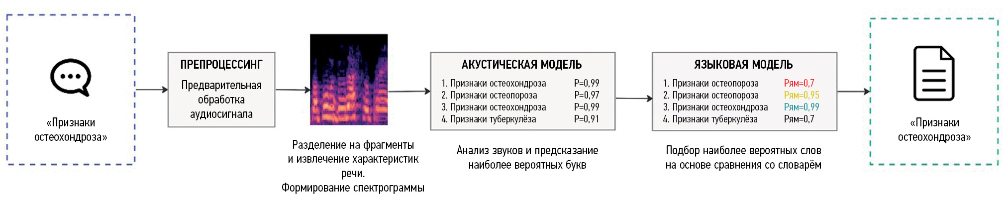
Speech recognition devices are promising tools for the healthcare system. Speech recognition technology has had a relatively long history of use in Western healthcare systems since the 1970s. However, it became widely used at the beginning of the 21st century, replacing medical transcriptionists. This technology is relatively new in home healthcare. Its active development began only in the early 2010s, and its implementation in healthcare started in late 2010. This delay is due to the idiosyncrasies of the Russian language and the limited computational power present at the beginning of the 21st century.
Currently, complexes of devices and software for speech recognition are used in the voice filling of medical records and can reduce the time for preparing reports for radiological examinations compared with traditional (keyboard) text input.
The literature review provides a brief history of speech recognition technology development and application in radiography. Key scientific studies showing its efficacy in Western healthcare systems are reflected. Voice recognition technology in the home is demonstrated, and its effectiveness is evaluated. The prospects for further development of this technology in Russian healthcare are described.
185-196
The role of dual-energy computed tomography in the diagnosis of gout and other crystalline arthropathies: A review
Abstract
The importance of dual-energy computed tomography in the diagnosis of gout, the principles of dual-energy computed tomography, the accuracy of the methodology, and the types of artifacts are discussed in this study. The possible dependence of the presence of deposits of sodium monourate on other clinical data and the role of dual-energy computed tomography in the differential diagnosis of other crystalline arthropathies are considered.
The dual-energy computed tomography has several advantages, including noninvasiveness, speed of execution, and a significant reduction in the risk of iatrogenic consequences compared with diagnostic arthrocentesis, which is the gold standard in diagnosing gout. Dual-energy computed tomography can accomplish gout detection, treatment, and differential diagnosis.
197-213
Case reports
Radiation methods in the diagnosis of primary and recurrent malignant ovarian struma: A case report
Abstract
We provide a rare clinical and diagnostic observation of primary and recurring malignant ovarian struma.
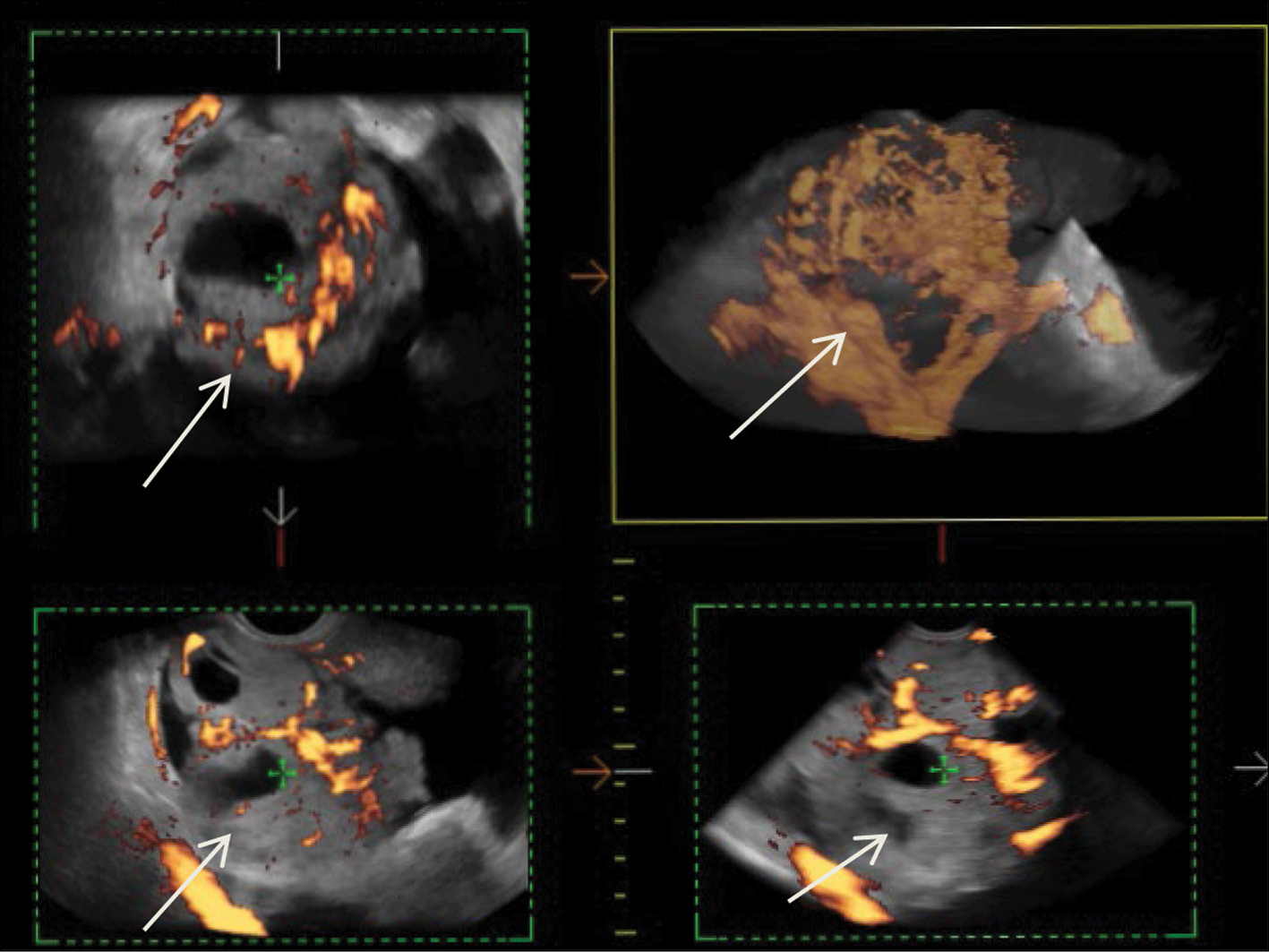
Malignant struma of the right ovary was detected 2 years after surgical treatment of primary benign struma of the left ovary. Six months later, the patient was diagnosed with a disease relapse, visualized exclusively according to radioisotope research methods. In the fourth year of anticancer treatment, ultrasonography revealed recurring foci along the peritoneum. According to the ultrasound data on the pelvic peritoneum and the projection of the removed right ovary, multiple solid nodes with high blood flow were visualized. Peak systolic velocity ranged from 2 to 9 cm/s in minor lesions from 4 to 12 mm, with an RI max of 0.53. For 4 years, the patient underwent radioiodine therapy with 131I with an activity of 6.0 GBq; the patient’s condition during the treatment was satisfactory.
214-225
Abernethy malformation: A case report
Abstract
Congenital portosystemic shunts are rare congenital vascular malformations characterized by a partial or complete portal blood diversion into the systemic circulation. Congenital extrahepatic portosystemic shunts, known as Abernethy malformations, pose a diagnostic challenge due to their low incidence and clinical presentations.
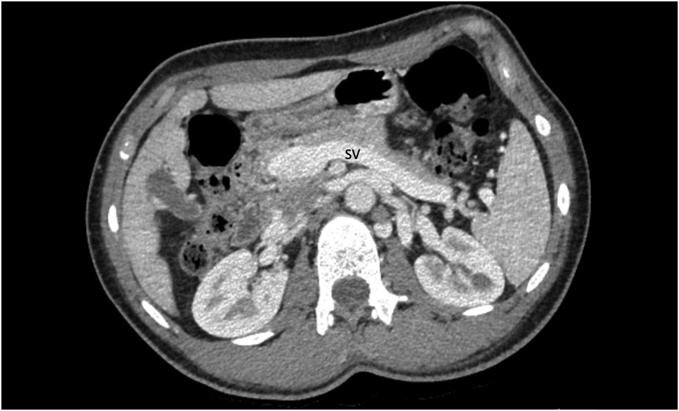
A 15-year-old male with a history of chronic epigastric pain and nausea, high arterial blood pressure, recurrent nose bleeds, chest pain, dizziness, dyspnea, low exercise tolerance, hematochezia, and itching was diagnosed with Abernethy malformation type Ib. Imaging studies revealed a dilated portal vein conduit flowing into the inferior vena cava, bypassing the porta hepatis. Multiple liver nodules, heart chamber dilatation, myocardial hypertrophy, and pulmonary hypertension were also discovered. Following multidisciplinary panel meetings, liver transplantation was advised due to the severity of the patient’s symptoms and shunt anatomy.
Furthermore, diagnostic algorithms and other treatment options are discussed.
226-237
Editorials
Hypothesis testing using R
Abstract
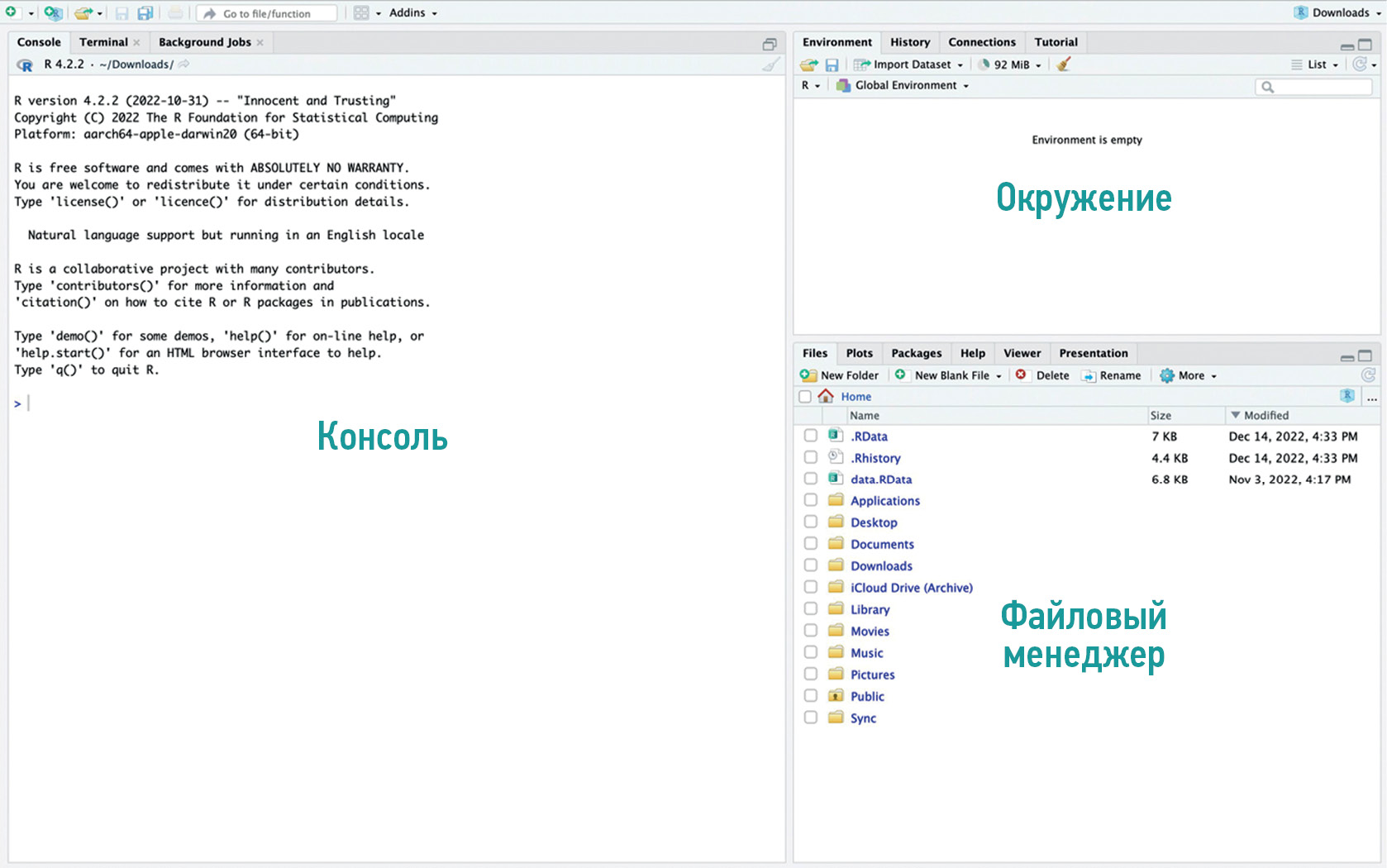
Competencies in statistical data processing are becoming increasingly important for modern scientists. The apparent advantages of open-source software for statistical analysis are its accessibility and adaptability. The programming language and the corresponding software R, available as a minimalistic console interface or a complete development environment RStudio/Posit, have the widest possibilities among free solutions.
We present a practical guide for comparing two groups using the software R. This study compares the effective doses of standard computed tomography with low-dose computed tomography for COVID-19 patients. The practical guide summarizes theoretical approaches to medical data processing and recommendations for correctly formulating research tasks and selecting optimal statistical analysis methods.
The main goal of the practical guide is to introduce the reader to the Posit interface and the basic functionality of the R language by using a practical example of treating a real medical problem. The presented material can be useful as an introduction to statistical analysis using the programming language R.
238-247
Регистрационный номер и дата принятия решения о регистрации СМИ: серия ПИ № ФС 77 - 79539 от 09 ноября 2020 г.